


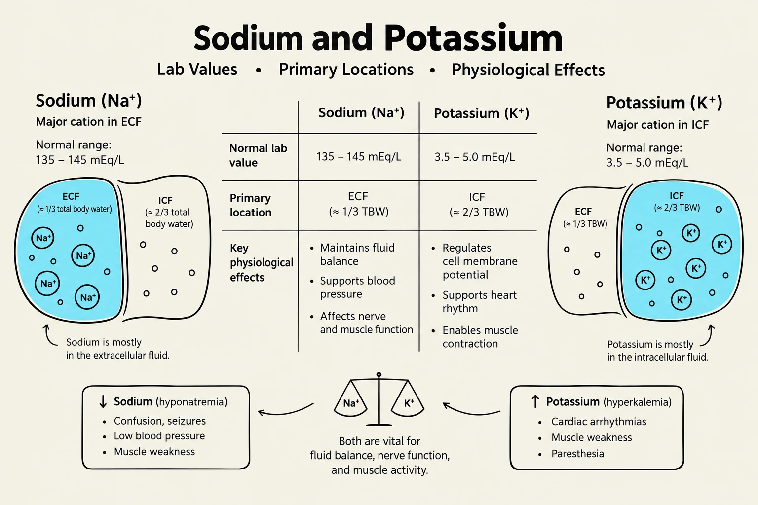
Maintaining homeostasis in the human body requires a delicate balance between water and solutes across various cellular compartments. This balance is not static; it is a dynamic equilibrium governed by physical forces and hormonal regulation. Understanding how fluids shift between the intracellular fluid (ICF) and extracellular fluid (ECF) compartments, and how electrolytes like sodium and potassium influence these shifts, is fundamental to clinical practice and nursing care.
The Mechanics of Fluid Movement: Starling Forces
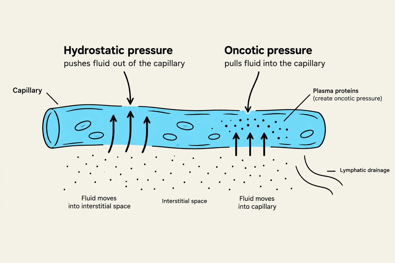
Fluid movement between the capillaries and the surrounding interstitial space is driven by Starling forces. Two primary pressures dictate this movement:
- Hydrostatic Pressure: This is the "pushing" force exerted by blood against the vessel walls. High hydrostatic pressure (often seen in heart failure) pushes fluid out of the capillaries and into the tissues, potentially causing edema.
- Oncotic Pressure: This is the "pulling" force exerted by plasma proteins, primarily albumin. Albumin acts like a sponge, pulling fluid back into the capillaries from the interstitial space.
When capillary permeability increases—due to inflammation, burns, or allergies—the vessel walls become "leaky," allowing proteins and fluid to escape more easily, further disrupting the balance.
Tonicity and Fluid Shifts
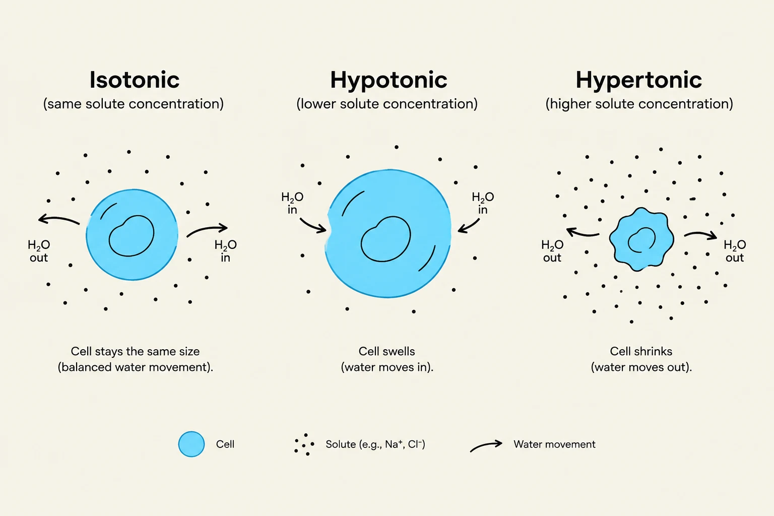
The movement of water into or out of cells is determined by the tonicity of the extracellular fluid. Because "water follows sodium," the concentration of solutes in the ECF dictates the osmotic gradient:
- Isotonic Fluids: The solute concentration is equal inside and outside the cell. Water moves in and out at equal rates, and the cell volume remains stable. Examples include 0.9% Normal Saline and Lactated Ringer's.
- Hypotonic Fluids: The ECF has a lower solute concentration than the cell. Water moves into the cells, causing them to swell.
- Hypertonic Fluids: The ECF has a higher solute concentration than the cell. Water is pulled out of the cells and into the vessels, causing the cells to shrink.
Keep reading for free.
Create an account to finish this reading and make it part of your study routine.
Continue reading freeAlready have an account? Log in
Updated