


The human immune system is a sophisticated network designed to distinguish "self" from "non-self." However, when this distinction fails, the resulting dysregulation can lead to devastating consequences, ranging from localized skin irritation to systemic organ failure. Understanding the pathophysiology of these disorders requires a deep dive into the mechanisms of hypersensitivity, the drivers of autoimmune inflammation, and the specific cellular pathways that govern dermatological and immune health.
Hypersensitivity: The Four Pillars of Immune Overreaction
Hypersensitivity refers to an exaggerated or inappropriate immune response to an antigen. These reactions are categorized into four distinct types based on the underlying mechanism and the speed of the response.
Type I: Immediate Hypersensitivity
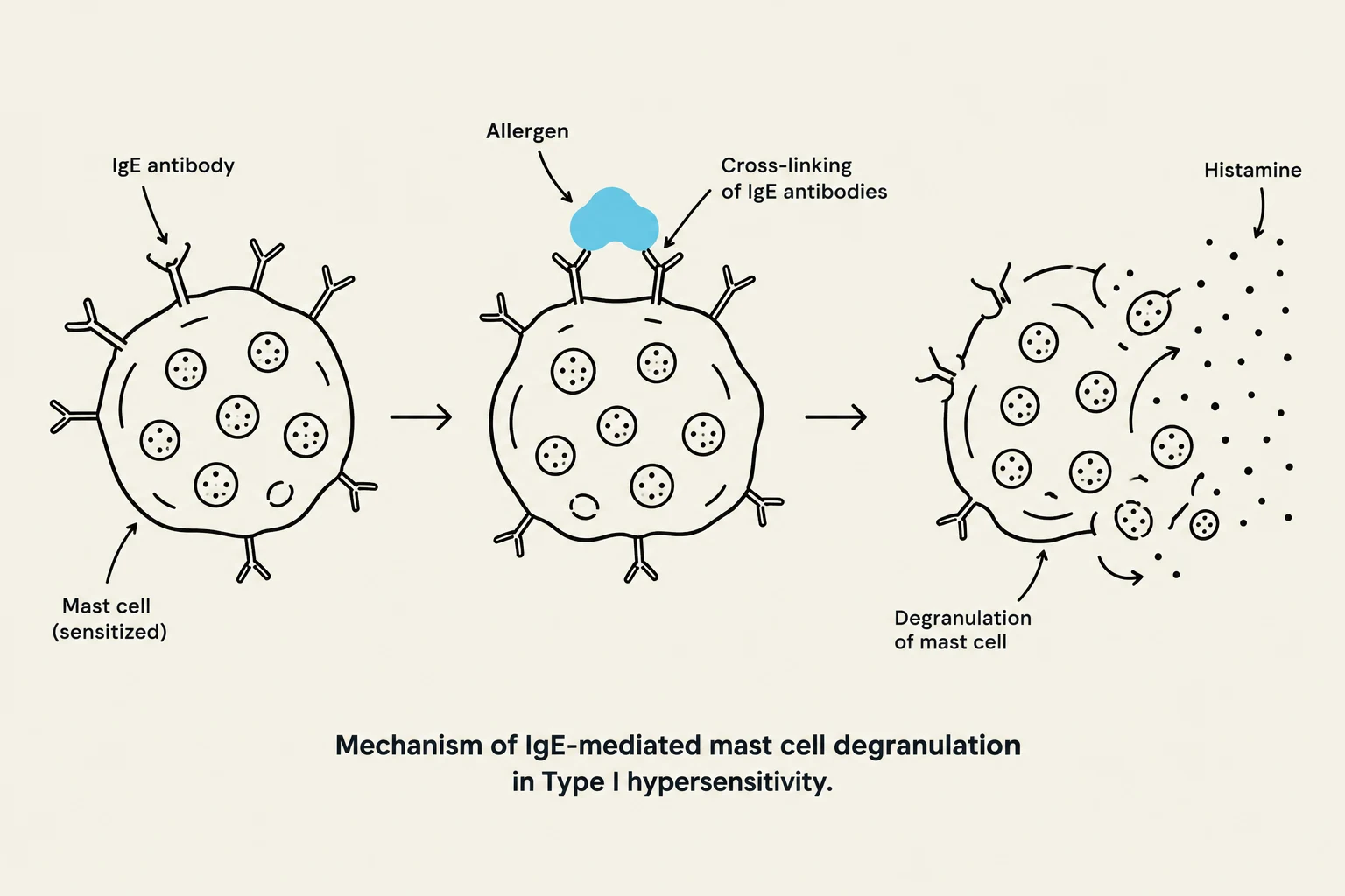
Type I reactions are IgE-mediated and occur almost immediately upon exposure to an allergen. When an individual is sensitized, IgE antibodies bind to mast cells. Upon re-exposure, the antigen cross-links these IgE antibodies, triggering mast cell degranulation and the release of inflammatory mediators like histamine. This process is responsible for symptoms ranging from localized hives to life-threatening anaphylaxis.
Type II: Cytotoxic Hypersensitivity
In Type II reactions, IgG or IgM antibodies target specific antigens located on the surface of the patient's own cells. This leads to cell destruction through complement activation or phagocytosis. A classic clinical example is hemolytic anemia, which can occur following a mismatched blood transfusion.
Type III: Immune Complex-Mediated Hypersensitivity
Type III hypersensitivity involves the formation of immune complexes—antigen-antibody clusters that circulate in the blood. These complexes eventually deposit in various tissues, such as blood vessel walls or kidney glomeruli, triggering complement activation and localized inflammation.

Keep reading for free.
Create an account to finish this reading and make it part of your study routine.
Continue reading freeAlready have an account? Log in
Updated