Chronic Kidney Disease (CKD): Definition and Risk Factors
Chronic Kidney Disease is defined as a progressive and persistent abnormality in kidney structure or function that lasts for ≥3 months. Diagnosis is based on two primary laboratory markers:
- An estimated GFR (eGFR) <60 mL/min/1.73 m2.
- The presence of albuminuria, typically defined as a Urine Albumin-to-Creatinine Ratio (UACR) >30 mg/g.
Major risk factors for the development of CKD include diabetes mellitus, hypertension, advanced age (>60 years), family history, and systemic inflammation.
The Pathophysiology of Progression
As nephrons are lost to injury, the remaining healthy nephrons undergo hypertrophy to compensate, a state known as hyperfiltration. To maintain GFR during this compensation, the body activates the renin-angiotensin-aldosterone system (RAAS), increasing Angiotensin II levels. While this maintains filtration temporarily, the resulting increase in intraglomerular pressure causes structural damage to the glomerulus, expanding the pores of the glomerular membrane. This allows proteins like albumin to leak into the urine (albuminuria), which further accelerates kidney damage and progressive nephron loss.
Drug-Induced Kidney Disease (DIKD)
Drug-Induced Kidney Disease refers to kidney injury resulting from medication exposure. It presents similarly to Acute Kidney Injury (AKI), characterized by a rise in serum creatinine (SCr) (≥0.3 mg/dL within 48 hours or ≥1.5X baseline within 7 days) and decreased urine output (<0.5 mL/kg/hr).
The pathophysiology of DIKD is categorized into three primary mechanisms:
1. Hemodynamic AKI (Functional Injury)
This involves changes in renal blood flow without direct structural damage to the tissue and is typically reversible.
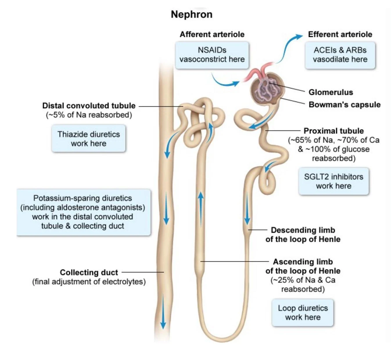
- Afferent Constriction: NSAIDs block prostaglandins, causing the afferent arteriole to constrict, reducing blood inflow.
- Efferent Dilation: ACE inhibitors (ACEIs) and ARBs block Angiotensin II, causing the efferent arteriole to dilate, which drops glomerular pressure.
- The "Triple Whammy": The simultaneous use of an ACEI/ARB, a diuretic, and an NSAID significantly increases the risk of hemodynamic AKI by crashing intraglomerular pressure.
2. Acute Tubular Necrosis (ATN) (Structural Injury)
ATN is the most common form of DIKD in inpatient settings. It involves direct toxicity to the tubular cells. As medications are concentrated in the tubules, cells die and slough off to form "muddy brown" casts, impairing reabsorption and filtration. Common nephrotoxins include aminoglycosides, vancomycin, amphotericin B, contrast media, and cisplatin.
3. Acute Interstitial Nephritis (AIN) (Immune-Mediated Injury)
AIN is an immune-mediated inflammatory response within the kidney interstitium. It is often triggered by drugs such as penicillins, sulfonamides, or PPIs. It may present with a classic triad of fever, rash, and eosinophilia, though this is often incomplete.
Exam Tip: Remember the distinction: Hemodynamic injury is functional (blood flow problem), whereas ATN and AIN are structural (actual tissue damage).
Pharmacologic Management of CKD Progression
Modern therapy focuses on slowing the decline of eGFR and reducing albuminuria through several key drug classes:
- RAAS Inhibitors (ACEIs and ARBs): These are recommended for all patients with albuminuria. By inhibiting Angiotensin II, they cause vasodilation of the efferent arteriole, which reduces intraglomerular pressure and protects the glomerulus from long-term damage. A modest rise in SCr (up to 30%) is expected and acceptable upon initiation.
- SGLT2 Inhibitors: These are considered kidney-protective drugs for patients with or without diabetes. They increase sodium delivery to the macula densa, leading to afferent arteriole constriction, which reduces glomerular pressure.
- Finerenone: A nonsteroidal mineralocorticoid receptor antagonist (MRA) that reduces aldosterone-mediated inflammation and fibrosis. It is particularly useful in patients with Type 2 Diabetes and CKD.
- GLP-1 Receptor Agonists: These are also utilized in the management of CKD, particularly in the context of diabetes.
Management of CKD Complications
As kidney function declines, several systemic complications arise:
Mineral and Bone Disorder (CKD-MBD)
When the kidneys cannot excrete excess phosphate, hyperphosphatemia occurs. Retained phosphate binds to calcium, lowering serum calcium levels. In response, the parathyroid gland releases parathyroid hormone (PTH) to pull calcium from the bones, leading to secondary hyperparathyroidism.
- Management: Includes dietary phosphate restriction, phosphate binders (taken with meals to bind dietary phosphate), Vitamin D analogs (to increase calcium absorption), and calcimimetics (to lower PTH in dialysis patients).
Hyperkalemia and Metabolic Acidosis
- Hyperkalemia: Managed by stabilizing the myocardium (calcium gluconate), shifting potassium intracellularly (insulin + dextrose), or removing potassium from the body (loop diuretics or potassium-binding resins like patiromer).
- Metabolic Acidosis: Caused by the impaired ability to regenerate bicarbonate. It is managed with oral sodium bicarbonate supplementation or dialysis.
Anemia of CKD
The kidneys are responsible for producing erythropoietin (EPO), the hormone that stimulates red blood cell production. Damaged kidneys produce less EPO, leading to a normocytic anemia.
- Management: The first step is ensuring adequate iron stores (ferritin and TSAT). If iron is sufficient but hemoglobin remains low (typically <10 g/dL), Erythropoiesis-Stimulating Agents (ESAs) are initiated.
Clinical Pearl: When treating anemia, you must correct iron levels first before initiating ESA therapy, as ESAs require iron to be effective.