An overview of cerebral vascular accidents (strokes), including their classification into ischemic and hemorrhagic types, underlying pathophysiology, and key risk factors.
A cerebral vascular accident (CVA), commonly known as a stroke or "brain attack," is a medical emergency that occurs when the blood supply to part of the brain is interrupted or when a blood vessel ruptures. Because the brain is a highly metabolic organ, consuming approximately 20% of the body's cardiac output (roughly 750–1,000 mL/min), it is extremely sensitive to changes in perfusion. Metabolic alterations begin within just 30 seconds of interrupted flow, and irreversible brain cell death can begin as early as the 5-minute mark.
Classification of Stroke
Strokes are primarily categorized into two types based on the underlying mechanism: ischemia or hemorrhage.
Ischemic Stroke (80–90% of cases)
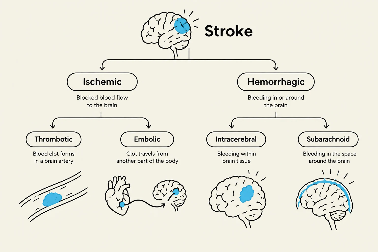
Ischemic strokes occur due to arterial occlusion, where blood flow is blocked by a physical obstruction. This is the most common type of stroke and is subdivided into:
Thrombotic Stroke: A blood clot (thrombus) forms locally within a cerebral artery. This is often the result of injury to the vessel wall or atherosclerosis, where plaque buildup narrows the lumen and creates a site for clot formation.
Embolic Stroke: A clot or debris forms elsewhere in the body—frequently in the heart due to conditions like atrial fibrillation, endocarditis, or heart valve disease—and travels through the bloodstream until it lodges in a cerebral artery.
Hemorrhagic Stroke
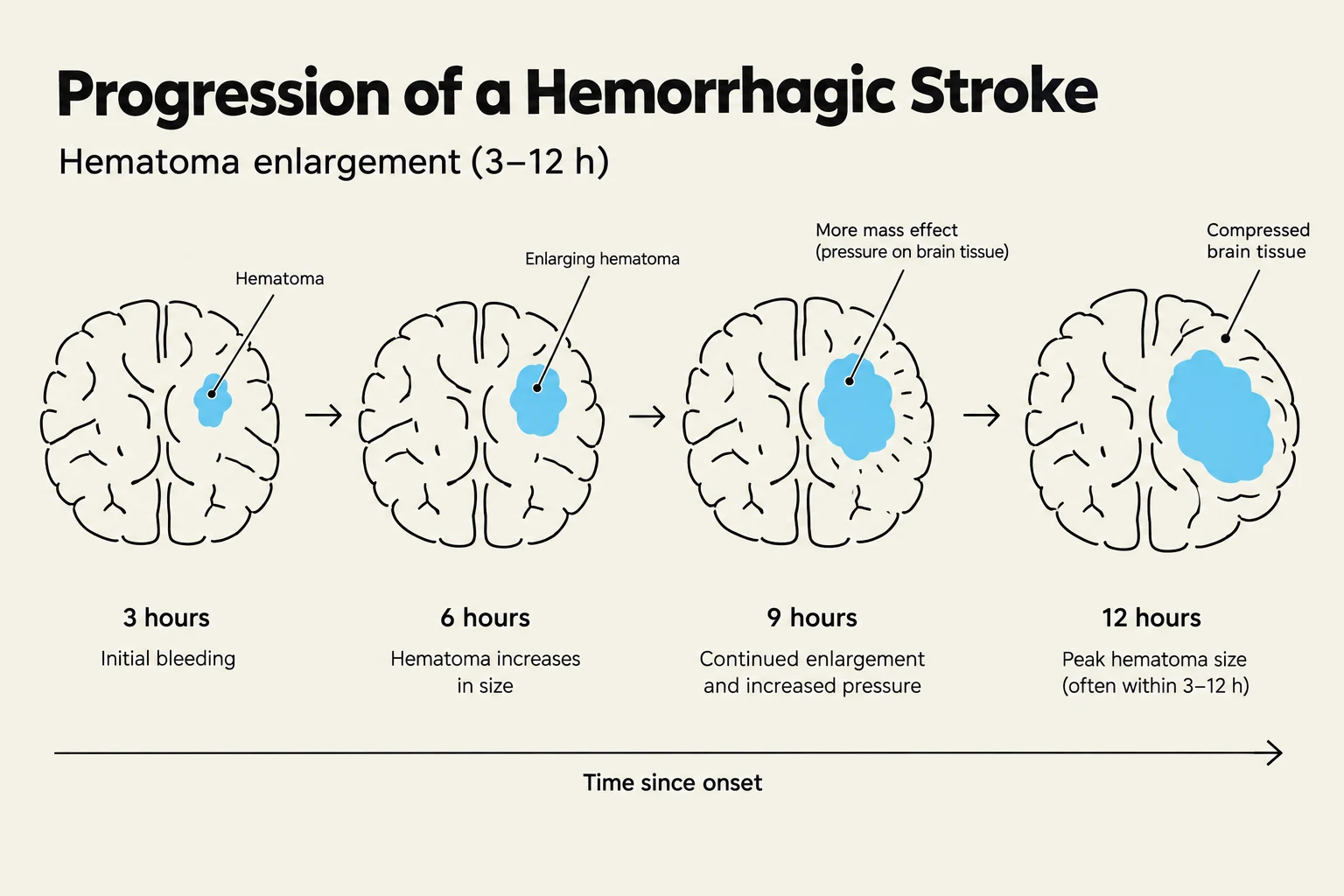
A hemorrhagic stroke occurs when a blood vessel ruptures, causing bleeding into or around the brain tissue. This bleeding increases intracranial pressure (ICP), which can compress brain tissue and decrease cerebral blood flow, leading to secondary injury.
Intracerebral Hemorrhage: Bleeding occurs directly within the actual brain parenchyma. The most common cause is chronic hypertension.
Subarachnoid Hemorrhage: Bleeding occurs in the subarachnoid space, between the brain and the meninges. This is most commonly caused by a ruptured aneurysm and is often characterized by a sudden, severe "thunderclap" headache.
This diagram illustrates the two main types of stroke: ischemic, caused by blocked blood flow, and hemorrhagic, caused by bleeding in or around the brain. Ischemic strokes are further divided into thrombotic (clot forms in an artery) and embolic (clot travels from elsewhere). Hemorrhagic strokes include intracerebral (bleeding within brain tissue) and subarachnoid (bleeding around the brain).
Pathophysiology and Compensation
The brain employs several mechanisms to maintain steady perfusion despite fluctuations in systemic conditions.
Autoregulation and Collateral Supply
Under normal conditions, the brain utilizes autoregulation to maintain constant blood flow by adjusting the diameter of its blood vessels. However, during an ischemic event, autoregulation becomes impaired, and the brain becomes increasingly dependent on systemic blood pressure to push blood past an occlusion.
Furthermore, carbon dioxide (CO2) levels act as a critical compensatory factor:
Decreased CO2: Causes vasoconstriction, which decreases cerebral blood flow.
Increased CO2: Causes vasodilation, which can increase blood flow.
To mitigate the effects of a blockage, the brain relies on collateral supply. The Circle of Willis is a vital anatomical structure that provides secondary pathways for blood flow, potentially bypassing narrowed or blocked main arteries to reduce the severity of ischemia.
Stroke in Evolution vs. Completed Stroke
Clinicians distinguish between a stroke in evolution, where symptoms are actively worsening over hours or days due to expanding infarction, cerebral edema, or fluctuating perfusion, and a completed stroke, where neurological deficits have stabilized and the brain has finished "evolving".
Risk Factors
Understanding risk factors is essential for both prevention and clinical assessment.
Modifiable Risk Factors
These are factors that can be managed or altered to reduce stroke risk:
Hypertension: The single highest risk factor. Chronic pressure damages vessel walls, leading to atherosclerosis or rupture.
Heart Disease: Conditions like atrial fibrillation or heart failure cause blood to pool abnormally, increasing clot formation risk.
Diabetes: High glucose levels damage vessel walls and increase blood viscosity.
Smoking: Nicotine causes vasoconstriction and vessel wall injury.
Obesity and Sleep Apnea: Obesity promotes inflammation and hypertension, while sleep apnea causes intermittent hypoxia.
Non-Modifiable Risk Factors
These are inherent characteristics that cannot be changed:
Age: Risk doubles every 10 years after age 50.
Sex and Ethnicity: It is more common in men, though women have higher mortality rates; African-Americans are twice as likely to experience a stroke.
History of TIA: A Transient Ischemic Attack (TIA) serves as a critical warning sign of worsening cardiovascular disease.
Clinical Presentation and Arterial Involvement
Symptoms of a stroke are highly dependent on which cerebral artery is affected.
Artery Involved
Common Clinical Presentations
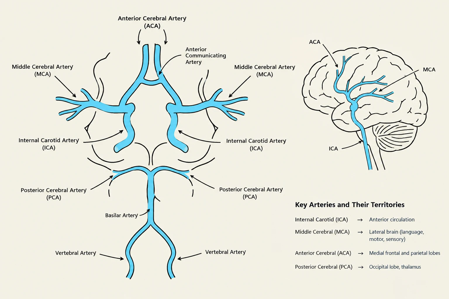
Middle Cerebral Artery (MCA)
Aphasia (difficulty speaking/understanding), dysphasia, visual field cuts, hemiparesis.
Vision field cuts, sensory impairment, dyslexia, coma, or blindness.
Vertebral-Basilar Artery
Dizziness (the #1 missed symptom), mouth/lip numbness, ataxia, slurred speech.
Diagram illustrating the major arteries supplying the brain, including the internal carotid, middle cerebral, anterior cerebral, posterior cerebral, vertebral, and basilar arteries, along with their respective territories.
Clinical Alert: When assessing a patient with potential brainstem involvement, the highest priority is monitoring for respiratory depression.
Management and Nursing Considerations
Management strategies vary significantly between ischemic and hemorrhagic strokes.
Ischemic Management
For ischemic strokes, the goal is to optimize perfusion. In some cases, clinicians must carefully manage blood pressure; while high blood pressure is a risk factor, in the presence of an acute stroke, a certain level of hypertension may be necessary to maintain perfusion past the blockage.
Hemorrhagic Management
For hemorrhagic strokes, the focus is on reducing intracranial pressure (ICP) and preventing complications like cerebral vasospasm (which typically peaks 6–10 days post-bleed).
Positioning: Elevate the head of the bed to 30∘ and keep the neck in a midline position to facilitate venous drainage and prevent jugular compression.
Fluid Management: Use isotonic solutions; avoid hypotonic solutions as they can worsen cerebral edema.
Medication: Calcium channel blockers (like Nimodipine) may be used to prevent vasospasm.
Exam Tip: If a patient has a subarachnoid hemorrhage, always monitor for a "thunderclap headache"—often described by patients as the "worst headache of my life".
This illustration shows the progression of a hemorrhagic stroke, detailing the enlargement of a hematoma in the brain over a 12-hour period.
Keep reading for free.
Create an account to finish this reading and make it part of your study routine.