Pathology of Hematological and Esophageal Disorders
An overview of the pathophysiology, signs, symptoms, diagnostics, and complications of various blood cell disorders, malignancies, and esophageal motility problems.
Understanding the complexities of the human body requires a deep dive into how specific systems—such as the blood and the digestive tract—respond to injury, infection, and malignancy. This article explores the intricate roles of white blood cells, the devastating impact of hematological cancers and anemias, the life-threatening nature of coagulation crises, and the motility issues that affect the esophagus.
Hematological Cell Functions and Indicators
The immune system relies on a diverse array of white blood cells (WBCs) to identify and neutralize threats. A normal WBC count typically ranges between $4,000$ and 10,000/mcL. When a clinician performs a differential count, they are looking at the specific percentages of these cells to determine the nature of an underlying issue.
The White Blood Cell Arsenal
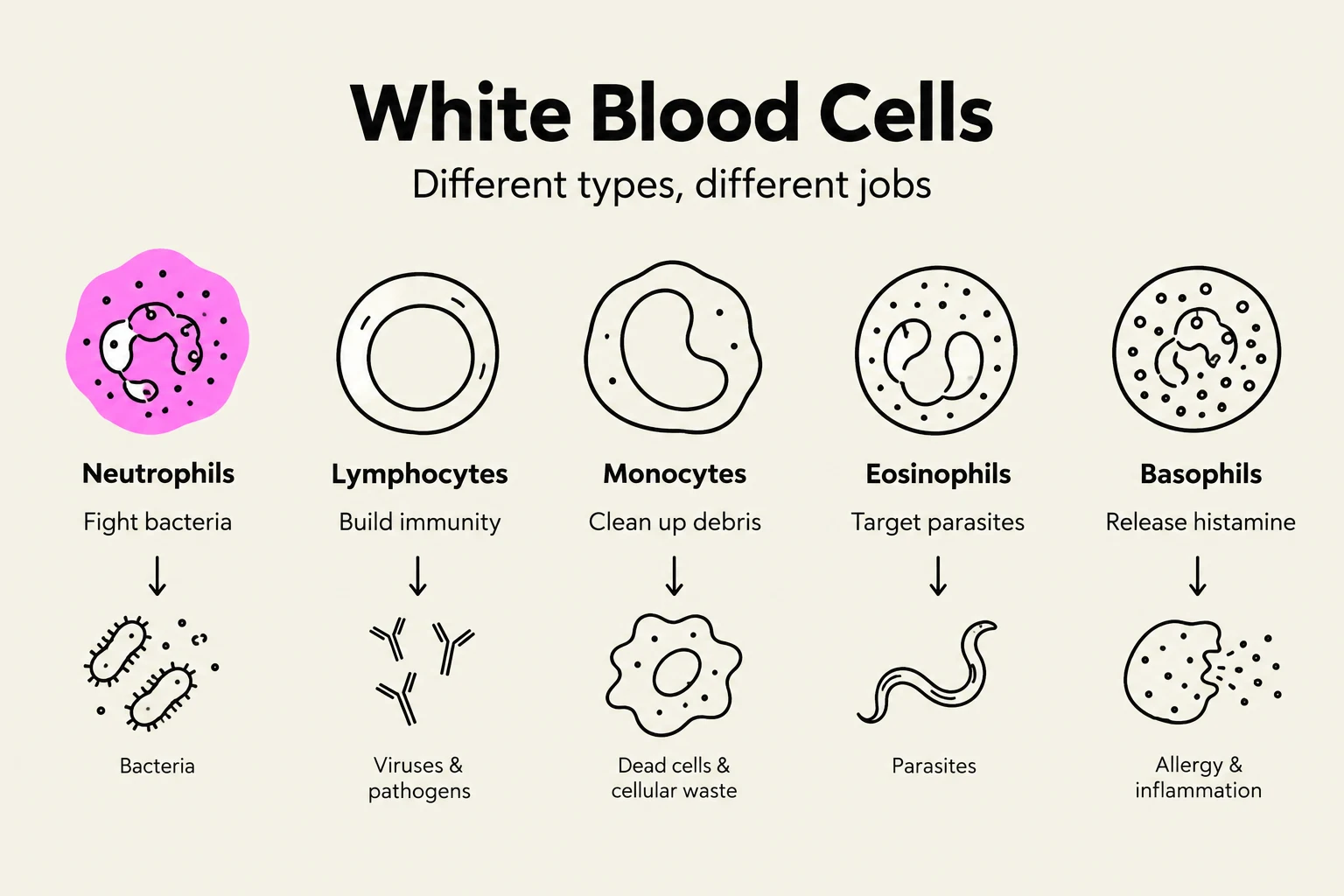
Neutrophils: These are the "first responders" to an infection, specifically targeting and destroying bacteria.
Lymphocytes: These cells provide the backbone of immune defense. B cells are responsible for producing antibodies, while T cells coordinate the immune response and directly destroy infected cells.
Monocytes: These cells act as scavengers, engulfing microorganisms and cellular debris; they can eventually differentiate into macrophages.
Eosinophils: These are specialized to fight parasitic infections (such as helminths) and play a significant role in allergic reactions and asthma.
Basophils: These cells release histamine and other chemicals that drive inflammation and allergic responses.
Exam Tip: The "Left Shift"
If a patient's lab results show a "left shift," it means there is an increased number of immature neutrophil bands in the blood. This is a critical clinical sign indicating that the bone marrow is working overtime to respond to a significant infection by releasing cells before they are fully mature.
This illustration depicts the five main types of white blood cells: neutrophils, lymphocytes, monocytes, eosinophils, and basophils, along with their respective functions and the targets they address in the body.
Hematological Malignancies
When the production of blood cells becomes unregulated, malignancies occur. These are broadly categorized into leukemias, lymphomas, and myelomas.
Leukemia
Leukemia involves the uncontrolled proliferation of abnormal blood cells. The specific type depends on the cell lineage involved:
Acute Lymphoblastic Leukemia (ALL): Characterized by dysfunctional stem cell precursors of B or T cells.
Chronic Lymphocytic Leukemia (CLL): Involves the proliferation of mature but functionally incompetent CD5+ B-cells.
Acute Myelogenous Leukemia (AML): Marked by the rapid proliferation of myeloid blast cells in the bone marrow.
Chronic Myelogenous Leukemia (CML): Involves the overproliferation of mature, non-functional myeloid cells.
Lymphoma and Multiple Myeloma
Lymphoma is a cancer of the lymphatic system. Hodgkin’s Lymphoma is specifically identified by the presence of Reed-Sternberg (RS) cells. In contrast, Non-Hodgkin’s Lymphoma involves the proliferation of abnormal B, T, or NK cells.
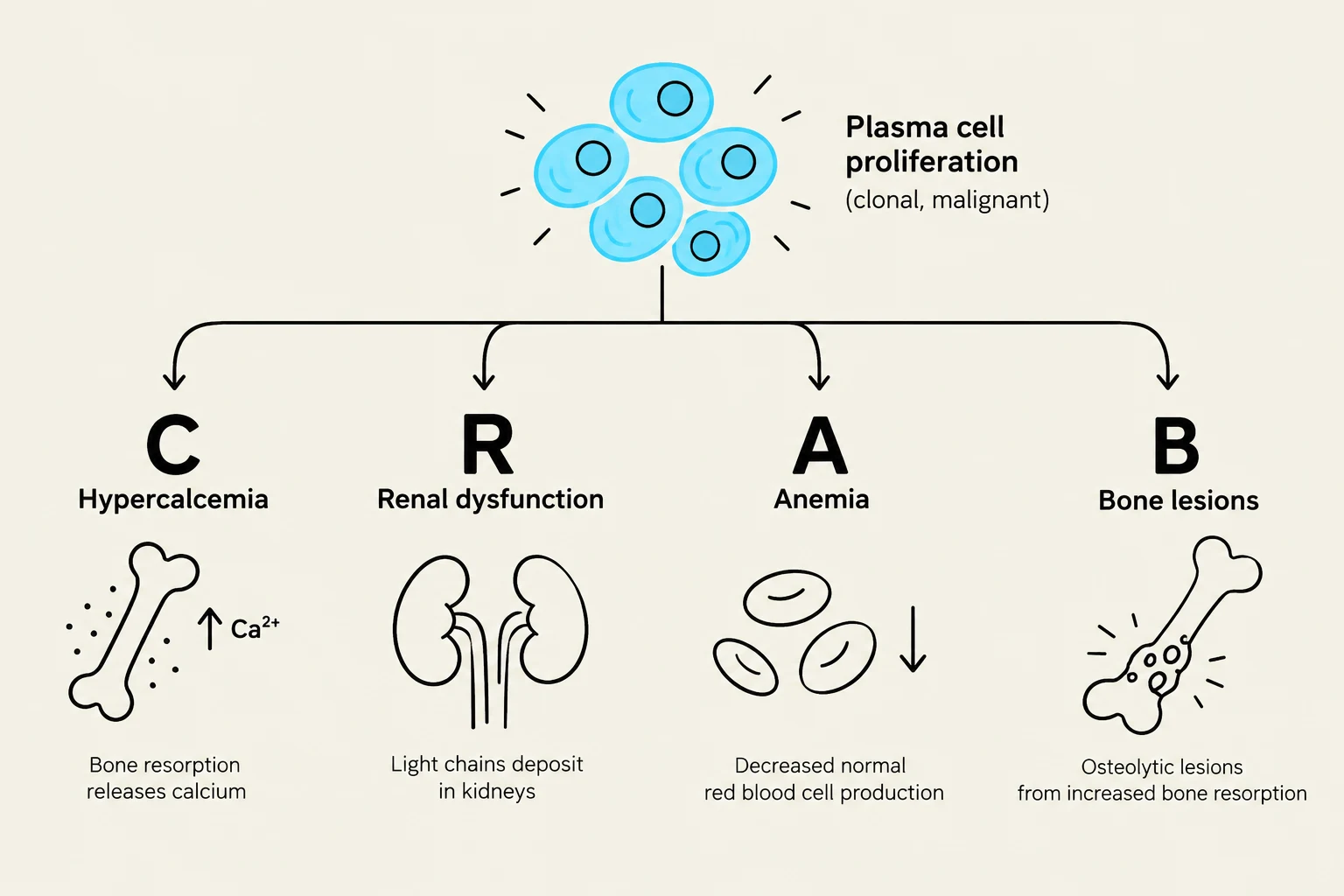
Multiple Myeloma is a neoplastic proliferation of plasma cells in the bone marrow. These cells produce excessive, abnormal immunoglobulins. Clinicians often use the CRAB mnemonic to remember its hallmark complications:
C: Calcium elevation
R: Renal dysfunction
A: Anemia
B: Bone deterioration
This diagram illustrates the CRAB mnemonic for the clinical manifestations of multiple myeloma: Hypercalcemia, Renal dysfunction, Anemia, and Bone lesions, all stemming from plasma cell proliferation.
Anemias and Coagulation Disorders
Anemias
Anemia is a deficiency in the blood's ability to carry oxygen, often due to low hemoglobin or red blood cell (RBC) counts.
Iron Deficiency Anemia (IDA): The most common form of anemia.
Pernicious Anemia: A specific type of Vitamin B12 deficiency caused by a lack of Intrinsic Factor (IF).
Aplastic Anemia: A severe condition where the bone marrow fails to produce all cell types, including RBCs, WBCs, and platelets.
Sickle Cell Anemia: A genetic hemoglobinopathy where red blood cells take on a sickle shape, leading to vaso-occlusive episodes and intense pain.
Coagulation and DIC
Bleeding disorders can be genetic, such as Hemophilia A (deficiency of Factor VIII) or Hemophilia B (deficiency of Factor IX).
More critically, Disseminated Intravascular Coagulation (DIC) is a secondary complication often triggered by sepsis, trauma, or cancer. In DIC, the clotting mechanism becomes overactivated, leading to a paradoxical state where the patient experiences simultaneous widespread clotting and severe bleeding, often accompanied by petechiae and hypotension.
Esophageal Motility and Disorders
The esophagus plays a vital role in transporting food to the stomach, but motility issues can cause significant distress.
GERD and Barrett's Esophagus
Gastroesophageal Reflux Disease (GERD) occurs when the lower esophageal sphincter (LES) has decreased tone or dysfunction, allowing acidic gastric contents to reflux into the esophagus. This can cause heartburn, dysphagia, and regurgitation.
If GERD is chronic, the constant acid exposure can lead to Barrett’s Esophagus, a metaplastic cellular change at the gastroesophageal junction. This is considered a precancerous change.
Achalasia and Hiatal Hernia
Achalasia is a motility problem involving the smooth muscle of the esophagus. It is characterized by increased muscular tone, a lack of peristalsis, and the incomplete relaxation of the LES, making patients feel as though food is getting "stuck".
Hiatal Hernia occurs when a portion of the stomach pushes upward through the diaphragm into the thoracic cavity, which can contribute to symptoms like substernal burning and belching.
Diagrams illustrating the differences between Achalasia, characterized by a lack of peristalsis and a tight Lower Esophageal Sphincter (LES), and GERD (Gastroesophageal Reflux Disease), characterized by a weak/incompetent LES leading to acid reflux.
Keep reading for free.
Create an account to finish this reading and make it part of your study routine.