


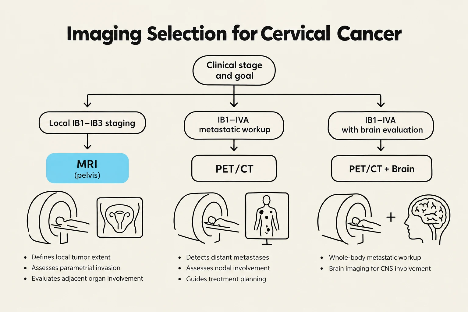
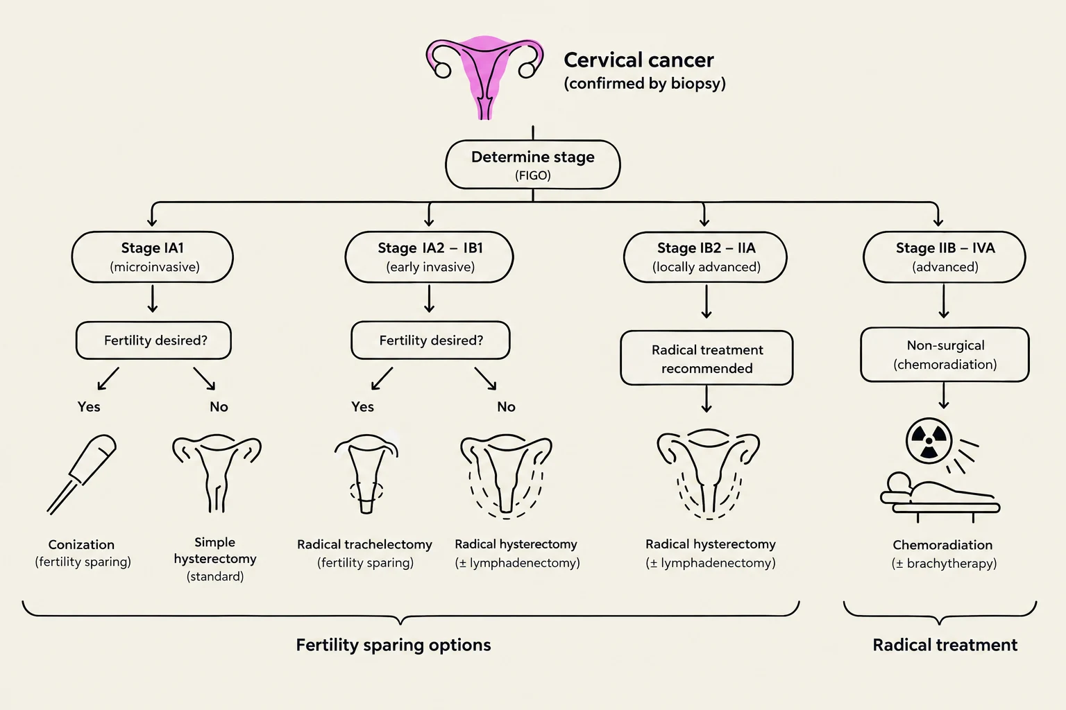
Cervical cancer management is a complex multidisciplinary effort that relies heavily on precise histological classification and accurate staging through imaging. Understanding the nuances between different tumor types—such as the distinction between common squamous cell carcinomas and rare neuroendocrine carcinomas—is critical for determining whether a patient should undergo conservative fertility-sparing surgery or more aggressive radical interventions. This article explores the pathological markers, imaging protocols, and clinical management strategies used in modern oncology.
Pathological Principles and Histology
The diagnosis of cervical cancer begins with a detailed microscopic examination of the tissue. While squamous cell carcinoma is the most common type, other histological subtypes present unique diagnostic challenges and clinical behaviors.
Adenosquamous Carcinoma
Adenosquamous carcinoma is an epithelial tumor characterized by the presence of both malignant squamous and glandular components. This subtype accounts for approximately 5%–6% of cervical cancer cases. Because it contains two distinct cell types, pathologists utilize specific immunohistochemistry (IHC) markers to differentiate them:
- Glandular components are highlighted using CK7, CEA, and PAX8.
- Squamous components are identified via p63 and p40.
- p16 overexpression is a hallmark of HPV-associated disease and is typically seen in both the squamous and glandular elements of this tumor.
Neuroendocrine Carcinoma (NECC)
Neuroendocrine carcinoma is a rare (<5%) but highly aggressive malignancy. It is frequently associated with HPV types 16 and 18. NECC is further categorized by cell morphology:
- Small Cell NECC: Characterized by hyperchromatic nuclei, scant cytoplasm, and high mitotic activity.
- Large Cell NECC: A distinct morphological variant within the neuroendocrine spectrum.
The diagnosis of NECC relies heavily on IHC markers. CD56 and synaptophysin are considered the most sensitive markers, whereas chromogranin provides the highest specificity. INSM1 is also a useful tool in the diagnostic toolkit. While TTF-1 positivity may be observed, it cannot be used to distinguish a primary cervical NECC from a metastasis originating in the lungs.
Keep reading for free.
Create an account to finish this reading and make it part of your study routine.
Continue reading freeAlready have an account? Log in
Updated