An explanation of the pancreas's role in digestion and blood sugar control, detailing insulin and glucagon functions, the incretin effect, and clinical monitoring of glucose levels.
The human body relies on a precise internal balance to maintain energy levels and cellular health. At the center of this metabolic control is the pancreas, a dual-purpose organ that manages both digestion and the complex orchestration of blood sugar levels. Understanding how the pancreas regulates glucose is fundamental to understanding metabolic health and the management of diabetes.
The Dual Role of the Pancreas
The pancreas performs two distinct primary functions: exocrine and endocrine. The exocrine function is dedicated to digestion, producing enzymes that help break down food in the small intestine. However, from a metabolic standpoint, the endocrine function is perhaps even more critical, as it involves the secretion of hormones directly into the bloodstream to regulate energy.
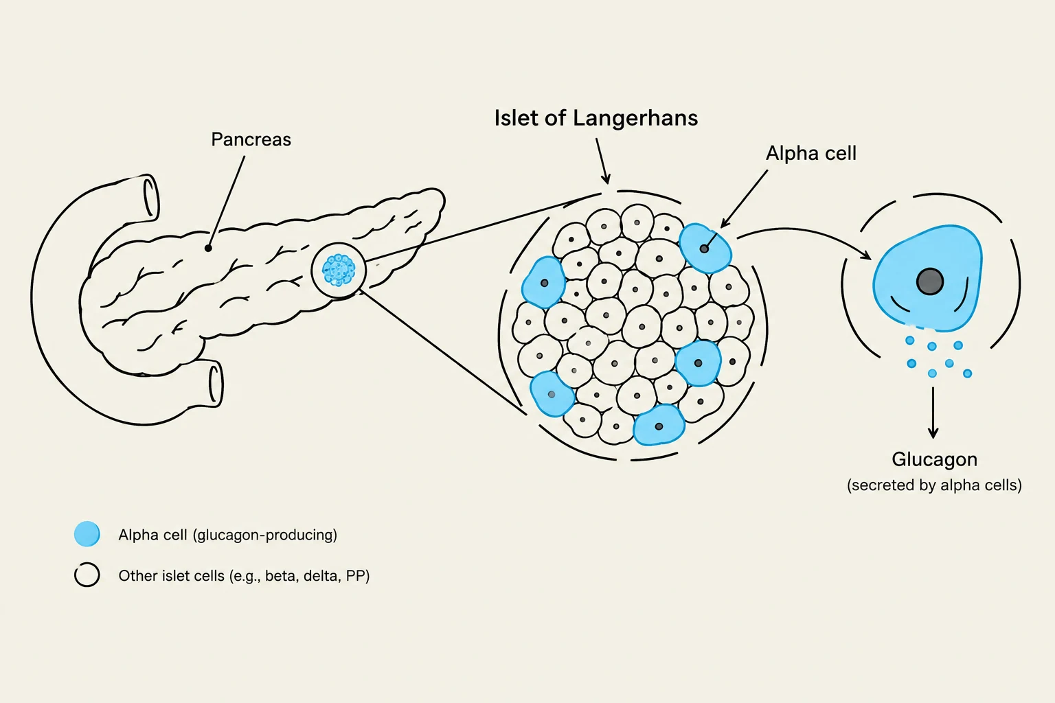
Within the endocrine system, specialized clusters of cells known as the islets of Langerhans act as the body's metabolic control center. These islets contain different cell types that respond to changing glucose levels:
Beta cells: These cells are responsible for producing insulin and amylin. Insulin is the primary hormone used to lower blood sugar.
Alpha cells: These cells secrete glucagon, a hormone that acts as the functional opposite of insulin.
Diagram illustrating the pancreas, Islets of Langerhans, alpha cells, and the secretion of glucagon.
Glucose Homeostasis: The Balancing Act
Glucose homeostasis is the continuous process of maintaining a stable concentration of glucose in the blood. This is achieved through a delicate balance between two forces: glucose uptake by the body's cells and glucose production by the liver.
The Role of Insulin
When you eat, your blood sugar rises. In response, the pancreatic beta cells release insulin. Think of insulin as a "key" that unlocks the cells of your body. Once the "key" opens the cell, glucose can enter to be used for energy, which subsequently lowers the concentration of sugar in the bloodstream.
The Role of Glucagon
Conversely, when blood sugar levels drop—such as during fasting, intense exercise, or the periods between meals—the alpha cells release glucagon. Glucagon signals the liver to undergo glycogenolysis, which is the mobilization of stored glycogen back into glucose. This process raises blood glucose levels back to a safe range.
Exam Tip: Remember that insulin and glucagon are antagonistic hormones. Insulin lowers blood glucose by promoting storage and uptake, while glucagon raises blood glucose by promoting release from the liver.
The Incretin Effect and the Role of DPP-4
The regulation of glucose is not just about the pancreas reacting to blood sugar; it also involves a sophisticated communication system from the gut known as the incretin effect.
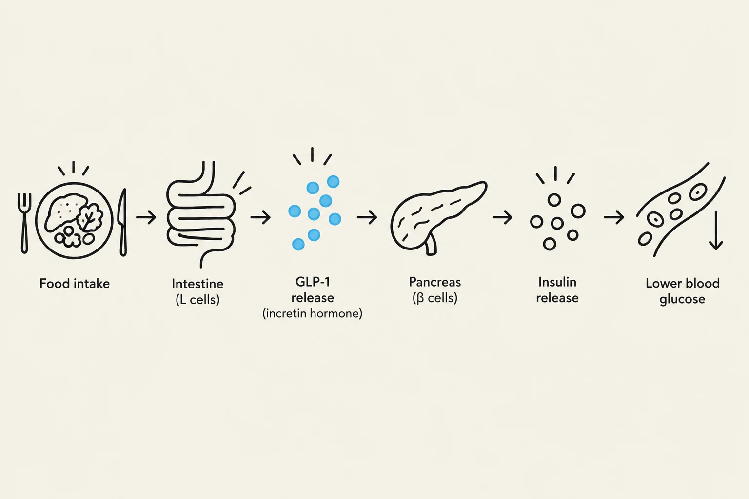
One of the most important incretins is Glucagon-like Peptide-1 (GLP-1). When food enters the system, GLP-1 is released and performs several vital tasks:
It signals the pancreas to increase insulin secretion (but only when glucose levels are high).
It decreases the secretion of glucagon.
It slows down gastric emptying (the rate at which the stomach empties), which helps prevent sudden spikes in blood sugar.
It promotes satiety, helping you feel full.
However, the body has a built-in "off switch" for this process. An enzyme called DPP-4 (dipeptidyl peptidase-4) rapidly destroys GLP-1, often within minutes of a meal.
To combat this rapid degradation, modern medicine utilizes two main pharmacological approaches:
GLP-1 Agonists: These drugs mimic the natural incretin (GLP-1) to stimulate insulin and promote weight loss.
DPP-4 Inhibitors: These drugs protect the body's natural GLP-1 from being destroyed by the enzyme, thereby prolonging the incretin effect.
Diagram illustrating the process of glucose regulation after food intake, involving the intestine, GLP-1 release, pancreas, and insulin release, leading to lower blood glucose levels.
Clinical Monitoring: FBG and HbA1c
To manage glucose effectively, clinicians monitor two primary metrics: Fasting Blood Glucose (FBG) and Hemoglobin A1c (HbA1c).
Fasting Blood Glucose (FBG)
FBG measures the glucose level in the blood after a period of fasting. The clinical interpretations are as follows:
Interpretation
Fasting Blood Glucose (mg/dL)
Normal
70–99 mg/dL
Prediabetes
100–125 mg/dL
Diabetes
≥126 mg/dL
Hemoglobin A1c (HbA1c)
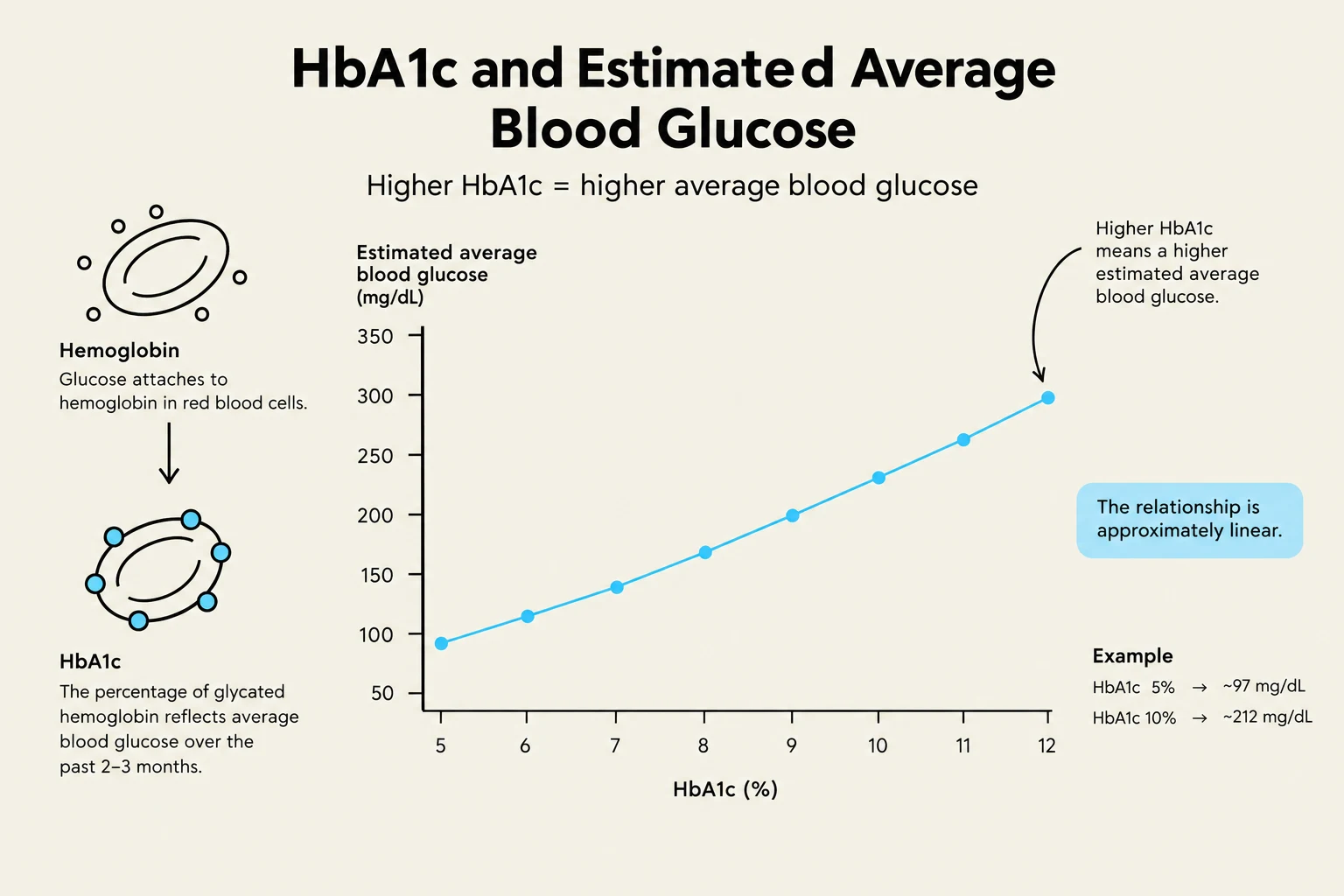
While FBG provides a "snapshot" of blood sugar at a single moment, HbA1c provides a long-term view. It measures the percentage of hemoglobin that has glucose attached to it, reflecting the average blood glucose levels over the approximately 120-day lifespan of a red blood cell.
There is a direct mathematical correlation between HbA1c and average blood glucose. For every $1%$ increase in HbA1c, the average blood glucose increases by approximately 30 mg/dL.
Worked Example:
If a patient has an HbA1c of $7%$, we can estimate their average glucose using the following relationship:
Average Glucose≈(HbA1c×30)+Baseline
Using the clinical correlation provided:
If HbA1c=7%→Average Glucose≈154 mg/dLIf HbA1c=8%→Average Glucose≈183 mg/dL
This graph illustrates the relationship between HbA1c levels and estimated average blood glucose. Higher HbA1c percentages correlate with higher average blood glucose levels, indicating a roughly linear relationship. The diagram also shows the process of glucose attaching to hemoglobin and defines HbA1c as a measure of average blood glucose over the past 2-3 months.
Clinical Note: Monitoring both values is essential. A patient might have a normal FBG but a high HbA1c, indicating that their blood sugar is spiking significantly after meals (postprandial hyperglycemia).
Keep reading for free.
Create an account to finish this reading and make it part of your study routine.