This content compares the oxygen binding dynamics of myoglobin and hemoglobin, explaining their structural differences, binding profiles (hyperbolic vs. sigmoidal), cooperativity, and physiological regulation through the Bohr effect and allosterism. It also touches upon the clinical relevance of hemoglobin structure in
In the study of biochemistry, understanding how proteins interact with ligands is fundamental to understanding life. One of the most elegant examples of this is the way respiratory proteins manage oxygen. While both myoglobin and hemoglobin serve the purpose of binding oxygen, they do so with vastly different dynamics, shaped by their unique molecular structures. These differences allow myoglobin to act as an efficient oxygen storage unit in muscles, while hemoglobin functions as a sophisticated delivery system that can sense and respond to the metabolic needs of tissues.
Structural Foundations: Monomers vs. Tetramers
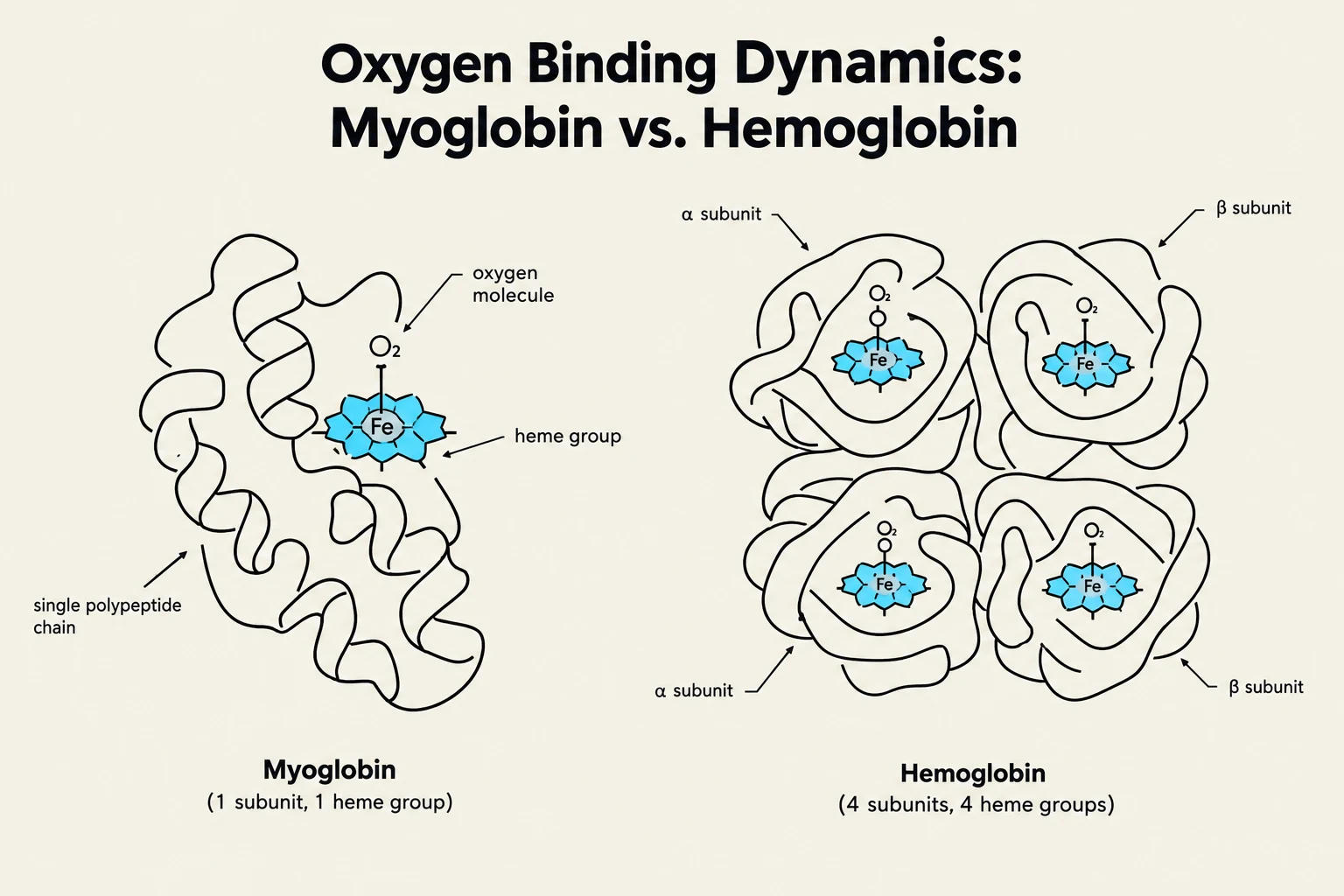
This diagram illustrates the structural differences between myoglobin and hemoglobin, highlighting their respective subunits and heme groups involved in oxygen binding.
The functional differences between these two proteins begin with their quaternary structure.
Myoglobin is a monomeric protein, meaning it consists of a single polypeptide chain. Within this chain is a single heme cofactor, which provides the specific site where molecular oxygen binds. Because it possesses only one binding site, the behavior of myoglobin is straightforward: it binds oxygen according to its affinity, but it cannot "communicate" with other molecules to change that affinity.
Hemoglobin, by contrast, is a heterotetrameric protein. It is composed of four subunits—typically two alpha (α) and two beta (β) subunits—that come together to form a complex quaternary structure. Each of these four subunits contains its own heme group, providing a total of four potential oxygen-binding sites. This multi-subunit arrangement is the prerequisite for the complex behavior known as cooperativity.
Binding Profiles: Hyperbolic vs. Sigmoidal
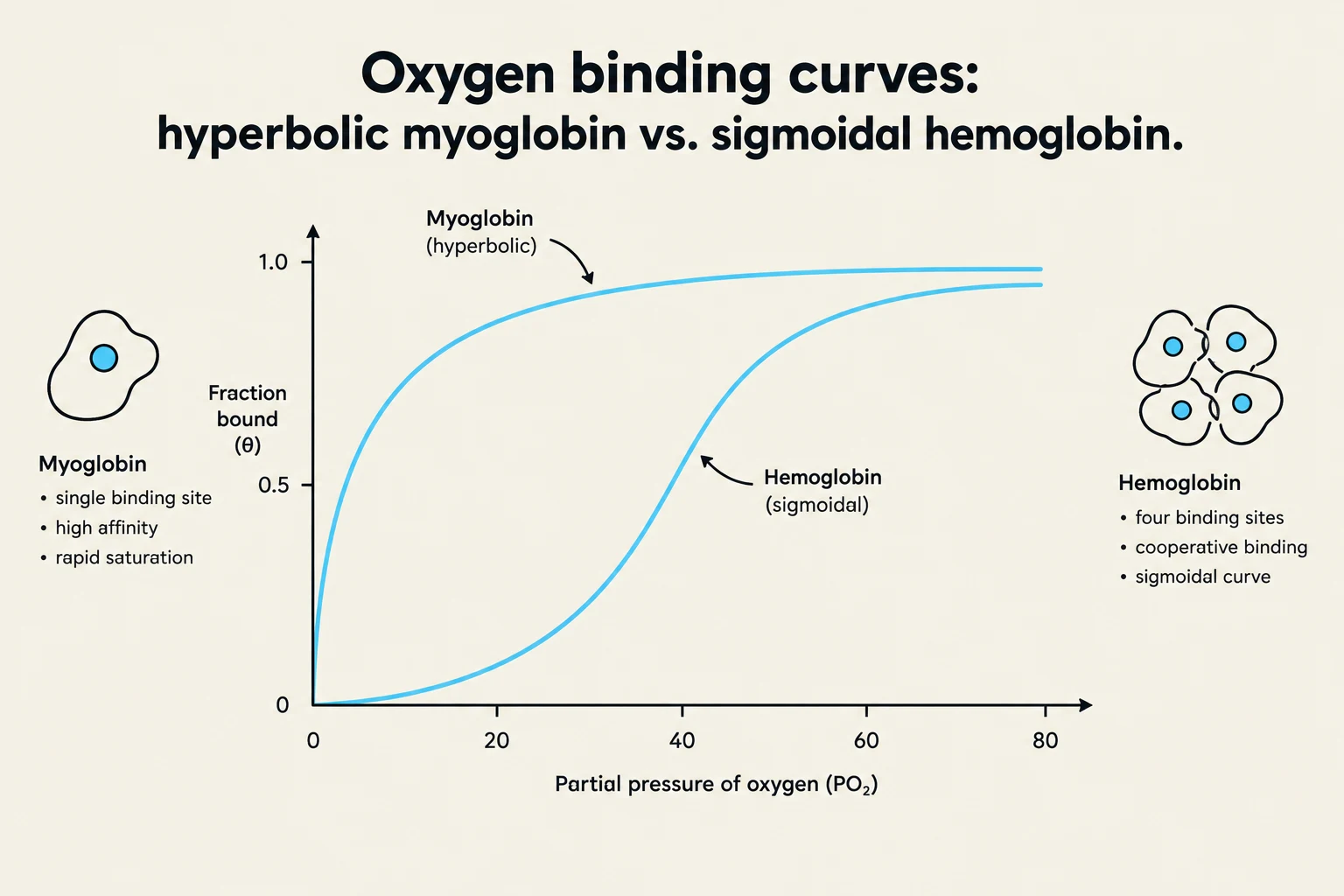
This graph illustrates the oxygen binding curves for myoglobin and hemoglobin, highlighting the hyperbolic curve of myoglobin and the sigmoidal curve of hemoglobin. It also details key characteristics of each protein, such as binding sites and affinity.
When we plot the fraction bound (θ) of oxygen against the partial pressure of oxygen (PO2), the resulting curves reveal the fundamental nature of their binding.
Myoglobin: The Hyperbolic Curve
Because myoglobin has only one site, its binding profile follows a hyperbolic curve. As the partial pressure of oxygen increases, the protein quickly reaches saturation. This profile is characteristic of non-cooperative binding, where the binding of one ligand molecule has no effect on the ability of the protein to bind subsequent molecules (since there is only one site to begin with).
Hemoglobin: The Sigmoidal Curve
Hemoglobin exhibits a sigmoidal (S-shaped) binding curve. This shape is the hallmark of positive cooperativity. In the early stages of oxygenation (at low partial pressures), the affinity for oxygen is relatively low. However, as more oxygen binds, the affinity of the remaining subunits increases significantly. This allows hemoglobin to be highly efficient: it binds oxygen tightly in the lungs (high PO2) and releases it readily in the tissues (low PO2).
Exam Tip: If you see a hyperbolic curve on a graph, think "monomer/non-cooperative" (Myoglobin). If you see an S-shaped curve, think "tetramer/cooperative" (Hemoglobin).
Quantifying Cooperativity: The Hill Coefficient
To mathematically describe the degree of cooperativity, scientists use the Hill coefficient (n). This value is an empirical constant that must be determined experimentally.
n=1: Represents non-cooperative binding. This is the case for myoglobin, where the binding of one oxygen molecule does not influence any other sites.
n>1: Represents positive cooperativity. For hemoglobin, the Hill coefficient is approximately 2.8.
Upper Bound: The value of n can never exceed the total number of binding sites. Therefore, for hemoglobin, n≤4.
The Linearized Hill Plot
Because fitting a sigmoidal curve to experimental data can be mathematically difficult, scientists use a linearization technique called a Hill plot. By plotting the ratio of the fraction bound to the fraction unbound against the logarithm of the partial pressure of oxygen, the relationship becomes linear:
log(1−θθ)=nlog(PO2)−log(Kd)
In this plot, the slope of the line represents the Hill coefficient (n). For hemoglobin, the plot typically shows two parallel lines at very low and very high oxygen concentrations, with a steep connecting line in the middle; the slope of that connecting line is the Hill constant.
Models of Cooperativity
How does the binding of oxygen to one subunit actually change the others? Two primary models attempt to explain this phenomenon:
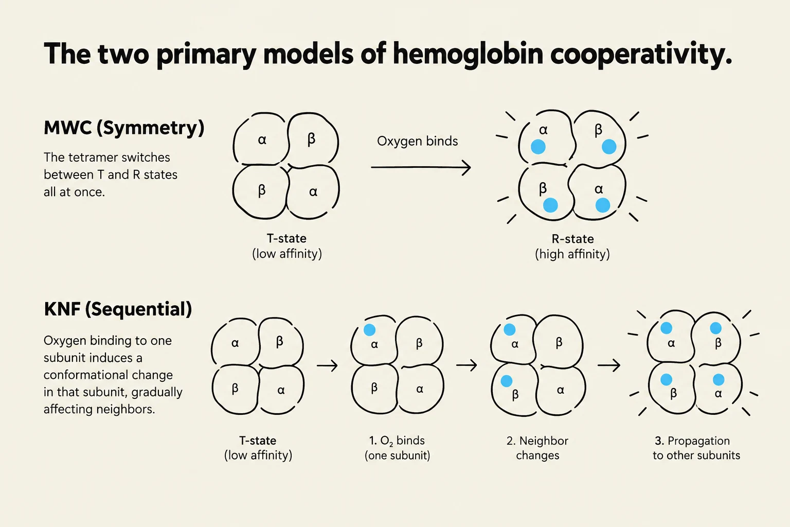
Symmetry (MWC) Model: This model proposes that the entire protein exists in an equilibrium between two distinct states: a T-state (Tense, low affinity) and an R-state (Relaxed, high affinity). The protein is symmetric; when oxygen binds, it shifts the equilibrium so that the entire tetramer transitions from the T-state to the R-state simultaneously.
Sequential (KNF) Model: This model suggests a step-by-step process. Oxygen binds to one subunit, inducing a conformational change in only that subunit. This change then influences the neighboring subunits, increasing their affinity slightly, but the protein does not transition all at once.
Current experimental evidence suggests that the reality lies somewhere between these two models, involving both subunit-specific changes and overall conformational shifts.
Physiological Regulation: The Bohr Effect and Allostery
Hemoglobin's ability to release oxygen is not just dependent on oxygen concentration, but also on the chemical environment of the tissue.
The Bohr Effect
The Bohr effect describes how a decrease in pH (increased acidity) reduces hemoglobin's affinity for oxygen. In metabolically active tissues, CO2 production increases. This CO2 reacts with water (catalyzed by carbonic anhydrase) to form carbonic acid, which dissociates into bicarbonate and protons ($H^+$), lowering the pH.
The increased concentration of protons leads to the protonation of specific amino acids, such as histidine. This protonation allows for the formation of new salt bridges (ionic interactions) that stabilize the T-state (the low-affinity, deoxy state). By stabilizing the T-state, the protein is encouraged to release its oxygen exactly where it is needed most.
Allosteric Regulation via 2,3-BPG
Another critical regulator is 2,3-bisphosphoglycerate (2,3-BPG), a metabolite from glycolysis. 2,3-BPG is a highly negatively charged molecule that binds to a specific pocket in the hemoglobin tetramer that only exists in the T-state. By binding to this pocket and forming salt bridges with positively charged residues (like lysine and arginine), 2,3-BPG stabilizes the T-state and promotes oxygen release. This is why individuals living at high altitudes, where oxygen is scarce, produce more 2,3-BPG to facilitate better oxygen delivery to tissues.
The image illustrates two models of hemoglobin cooperativity: the MWC (Symmetry) model where the tetramer switches between T and R states simultaneously, and the KNF (Sequential) model where oxygen binding to one subunit induces gradual conformational changes affecting neighbors.
Clinical Correlation: Sickle Cell Anemia
The importance of hemoglobin's structure is highlighted by sickle cell anemia. This disease is caused by a single point mutation in the β-subunit gene, where the polar amino acid glutamic acid is replaced by the non-polar amino acid valine at position six.
Because valine is hydrophobic, it creates a "sticky" patch on the surface of the protein that seeks to avoid water. This causes hemoglobin molecules to aggregate into long fibers when they are in the deoxygenated state, distorting the red blood cell into a "sickle" shape. This aggregation disrupts the normal conformational changes required for efficient oxygen binding and release.
Keep reading for free.
Create an account to finish this reading and make it part of your study routine.