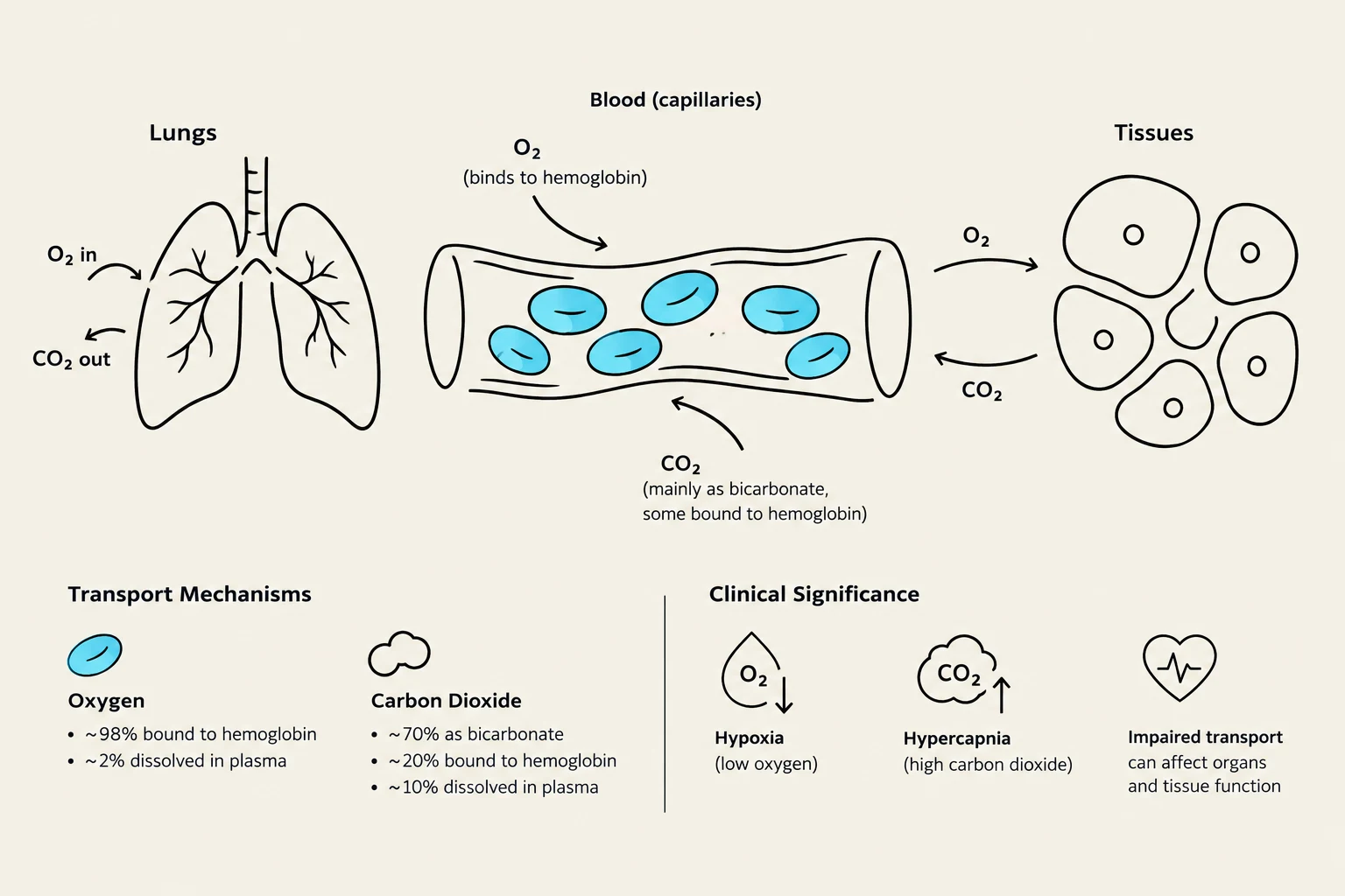
Hemoglobin (specifically Hb A in adults) is composed of two main parts:
- Four Heme Groups: These are the pigmented, nonprotein portions that contain iron. The iron is the specific site where oxygen binds.
- Four Globin Chains: These are the polypeptide (protein) chains that provide the structure for the heme groups.
When hemoglobin is bound to oxygen, it is referred to as oxyhemoglobin; when it is not bound, it is called reduced hemoglobin or deoxyhemoglobin.
Calculating Oxygen Content and Delivery
To understand a patient's oxygenation status, clinicians must distinguish between the pressure of the gas (PaO2) and the total content of oxygen in the blood (CaO2).
Arterial Oxygen Content (CaO2)
The total amount of oxygen in arterial blood is the sum of the oxygen dissolved in the plasma and the oxygen bound to hemoglobin. The formula to calculate this is:
CaO2=(Hb×1.34×SaO2)+(PaO2×0.003)
In this equation:
- Hb is the hemoglobin concentration (normal male: 14–16 g/dL; normal female: 12–15 g/dL).
- 1.34 mL is the amount of O2 carried by each gram of hemoglobin.
- SaO2 is the arterial hemoglobin saturation.
- PaO2×0.003 represents the dissolved portion.
A normal CaO2 is approximately 20 mL/dL.
Mixed Venous Oxygen Content (CvO2)
As blood travels through the tissues, oxygen is unloaded. The content of the mixed venous blood is calculated similarly:
CvO2=(Hb×1.34×SvO2)+(PvO2×0.003)
The normal value for CvO2 is approximately 15 mL/dL.
Total Oxygen Delivery (DO2)
The total amount of oxygen delivered to the tissues per minute depends on both the oxygen content and the volume of blood pumped by the heart (cardiac output, Qt):
DO2=Qt×(CaO2×10)
Clinical Note: Polycythemia
If a patient suffers from chronic low PaO2 (hypoxia), the body may attempt to compensate by producing more red blood cells. This condition, known as secondary polycythemia, results in an abnormally high hematocrit (>$52%$ in men, >$48%$ in women) and elevated hemoglobin levels.
The Oxyhemoglobin Dissociation Curve
The relationship between the partial pressure of oxygen (PO2) and the percentage of hemoglobin saturation is not linear; it is represented by an S-shaped (sigmoidal) curve.
The Flat Portion (The Safety Zone)
At normal arterial PO2 levels (80–100 mmHg), the curve is relatively flat. This serves as a loading safety zone in the lungs. Because the curve is flat here, even if the PaO2 drops slightly, the hemoglobin saturation (SaO2) remains relatively stable, ensuring the blood remains well-saturated before leaving the lungs.
The Steep Portion (The Unloading Zone)
When PaO2 falls below 80 mmHg (and especially below 60 mmHg), the curve enters its steep portion. In this range, small decreases in PO2 lead to massive decreases in hemoglobin saturation. This is physiologically advantageous for rapid oxygen unloading at the tissue level, where PO2 is naturally lower.
The P50 and Curve Shifts
The P50 is a critical constant representing the partial pressure of oxygen at which hemoglobin is $50%$ saturated. The normal P50 is approximately 27 mmHg. Changes in the chemical environment can shift the entire curve, altering the affinity of hemoglobin for oxygen.
Right Shift: "Right Releases"
A right shift means the P50 increases, indicating that hemoglobin has a decreased affinity for oxygen. This makes it easier for hemoglobin to release (unload) oxygen to the tissues.
- Causes: Increased temperature, increased PaCO2, increased 2,3-BPG, and acidosis (decreased pH). This relationship between pH and oxygen affinity is known as the Bohr effect.
- Clinical Associations: COPD, ketoacidosis, and chronic anemia.
- Impact: At a PaO2 of 60 torr, a right-shifted curve might result in an SaO2 of only ∼75% compared to the normal ∼90%.
Left Shift: "Left Loves"
A left shift means the P50 decreases, indicating that hemoglobin has an increased affinity for oxygen. Hemoglobin "loves" oxygen and holds onto it more tightly, making it harder to unload at the tissues.
- Causes: Decreased temperature, decreased PaCO2, decreased 2,3-BPG, alkalosis (increased pH), fetal hemoglobin (HbF), and carboxyhemoglobin (COHb).
- Clinical Associations: Hyperventilation and methemoglobinemia.
- Impact: To force oxygen dissociation in a left-shifted state, the tissue PO2 must drop much lower than normal. For example, at a pH of 7.6, PO2 might need to fall from 60 torr to 30 torr just to unload 5 vol% of oxygen.
Physiological Shunts and Monitoring
Even in healthy individuals, hemoglobin saturation is typically around $97%$ rather than $100%$ due to physiological shunts. These include:
- Anatomic Shunts: Such as the Thebesian veins (which drain directly into the heart) and bronchial veins.
- Capillary/Functional Shunts: Such as underventilated alveoli, where blood passes by areas of the lung that are not receiving adequate fresh air.
Clinical Monitoring: Pulse Oximetry
Clinicians monitor oxygenation using pulse oximetry (SpO2), which utilizes spectrophotometry (measuring light absorption) to estimate hemoglobin saturation.
Exam Tip: The 40-50-60 / 70-80-90 Rule
There is a predictable relationship between PaO2 and SaO2:
- PaO2≈40 mmHg→SaO2≈70%
- PaO2≈50 mmHg→SaO2≈80%
- PaO2≈60 mmHg→SaO2≈90%
Warning: Pulse oximeter readings below $70%$ saturation are often considered inaccurate and unreliable.