Explores the integumentary system, including the skin, hair, nails, and glands, detailing its functions in homeostasis, protection, thermoregulation, and Vitamin D synthesis. Covers skin layers (epidermis, dermis, hypodermis), accessory structures, sensory receptors, and clinical considerations like aging and injury.
The integumentary system is much more than just the outer covering of our bodies; it is a complex, multi-functional organ system essential for survival. It comprises the skin and its accessory structures, including hair, nails, oil (sebaceous) glands, sweat glands, and various sensory receptors. By performing a wide array of tasks—from protecting us against environmental hazards to regulating our internal temperature—the integumentary system plays a vital role in maintaining homeostasis, the stable internal environment required for the body to function correctly.
Functions and Homeostasis
The primary mission of the integumentary system is to maintain homeostasis. It achieves this through several specialized mechanisms:
Thermoregulation: The system helps the body maintain its temperature through the production of sweat and by adjusting blood flow to the dermis.
Protection: It acts as a barrier against physical injury, pathogens, and UV radiation using components like keratin, melanin, and acidic sweat.
Sensory Perception: Through various sensory receptors, the skin provides vital information about the external world.
Vitamin D Synthesis: One of its unique chemical functions is the conversion of inactive vitamin D precursors into their active form via ultraviolet (UV) rays. This active vitamin D is crucial for the absorption of calcium from the food we eat.
Other Roles: It also serves as a blood reservoir, an excretory organ, and a site for the absorption of certain materials.
Study Tip: When thinking about homeostasis, remember that the integumentary system is the body's "interface." It manages what comes in (absorption), what goes out (excretion/sweat), and how we react to the environment (sensory/temperature).
The Layers of the Skin
The skin is organized into distinct layers, each with a specific composition and function. While we often think of "skin" as a single unit, it is actually composed of two major layers: the epidermis and the dermis.
The Epidermis
The epidermis is the most superficial layer, meaning it is the one in direct contact with the outside world. It is composed of four major types of cells: keratinocytes, melanocytes, intraepidermal macrophages (Langerhans cells), and tactile epithelial cells (Merkel cells).
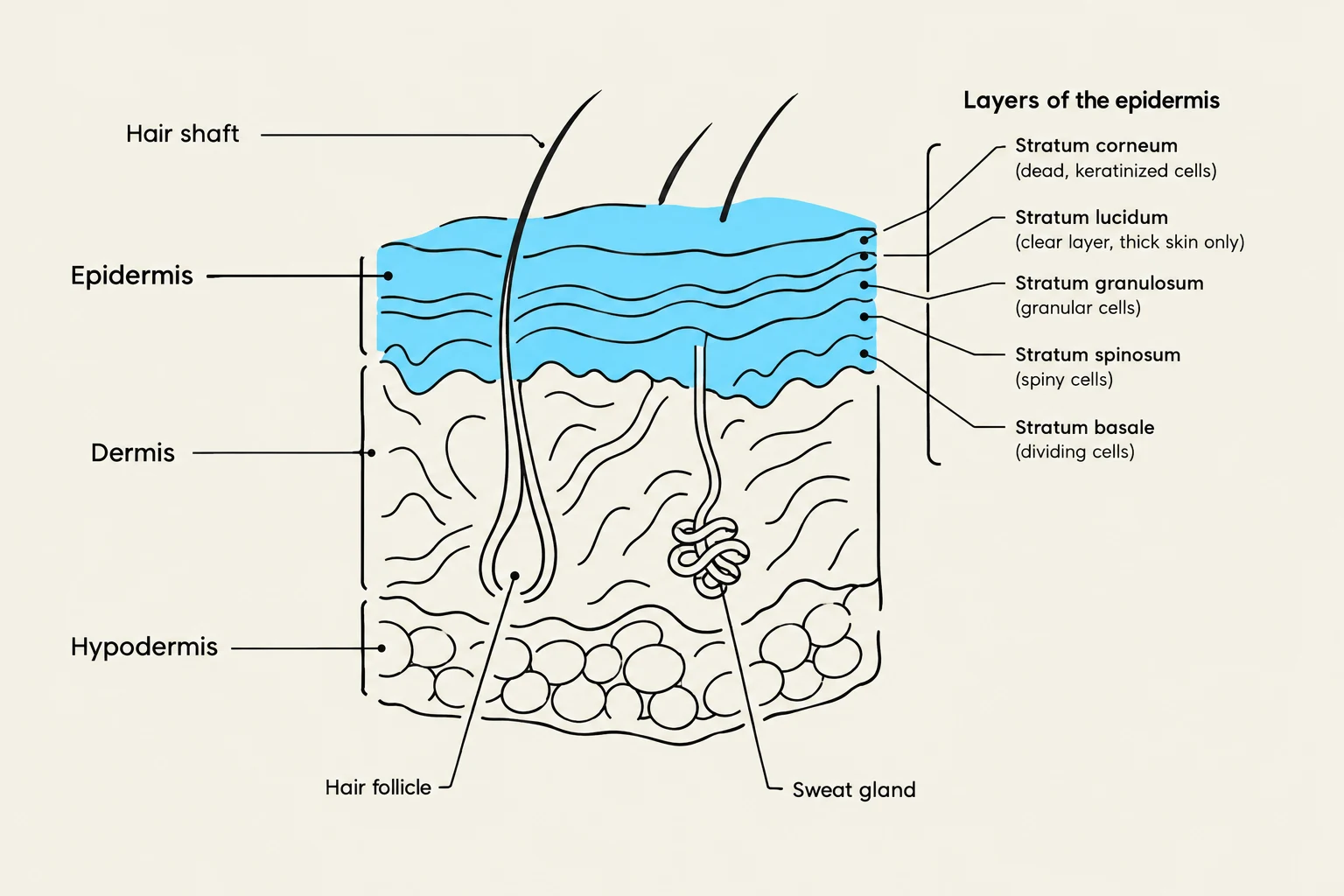
The thickness of the epidermis varies depending on the body region. "Thin skin" covers most of the body, while "thick skin"—which lacks hair—is found specifically on the palms of the hands and the soles of the feet. In thick skin, the epidermis contains five distinct layers (strata), whereas thin skin only contains four. From deepest to most superficial, these layers are:
Stratum basale
Stratum spinosum
Stratum granulosum
Stratum lucidum (present only in thick skin)
Stratum corneum
Diagram illustrating the layers of the epidermis, including the stratum corneum, stratum lucidum, stratum granulosum, stratum spinosum, and stratum basale, along with the hair follicle and sweat gland within the dermis and hypodermis.
The Dermis
Located directly beneath the epidermis is the dermis. Unlike the cellular epidermis, the dermis is composed of connective tissue that is rich in collagen and elastic fibers, providing the skin with strength and flexibility. The dermis is also highly vascular; it contains enough blood vessels to hold approximately 8–10% of the body's total blood volume while an adult is at rest.
The Hypodermis (Subcutaneous Layer)
Deep to the dermis lies the hypodermis, also frequently referred to as the subcutaneous layer. It is important to note that the hypodermis is not technically considered a layer of the skin, but rather a layer that attaches the skin to the underlying tissues and organs. It is primarily composed of areolar and adipose (fat) tissue.
Accessory Structures and Sensory Receptors
The integumentary system includes several "accessory structures" that emerge from the skin layers to perform specialized tasks.
Hair and Nails
Hair is composed of dead, keratinized epidermal cells and is found on most body surfaces except the palms and soles. Hair consists of a shaft (above the surface), a follicle (below the surface), and a root that penetrates into the dermis. Nails are also made of keratinized epidermal cells and serve to protect the digits.
Glands
There are four main types of glands associated with the skin:
Sebaceous (oil) glands: Connected to hair follicles.
Eccrine sweat glands: The most numerous type.
Apocrine sweat glands: Located mainly in hairy areas.
Ceruminous glands: Modified sweat glands found in the ear canal.
Sensory Receptors
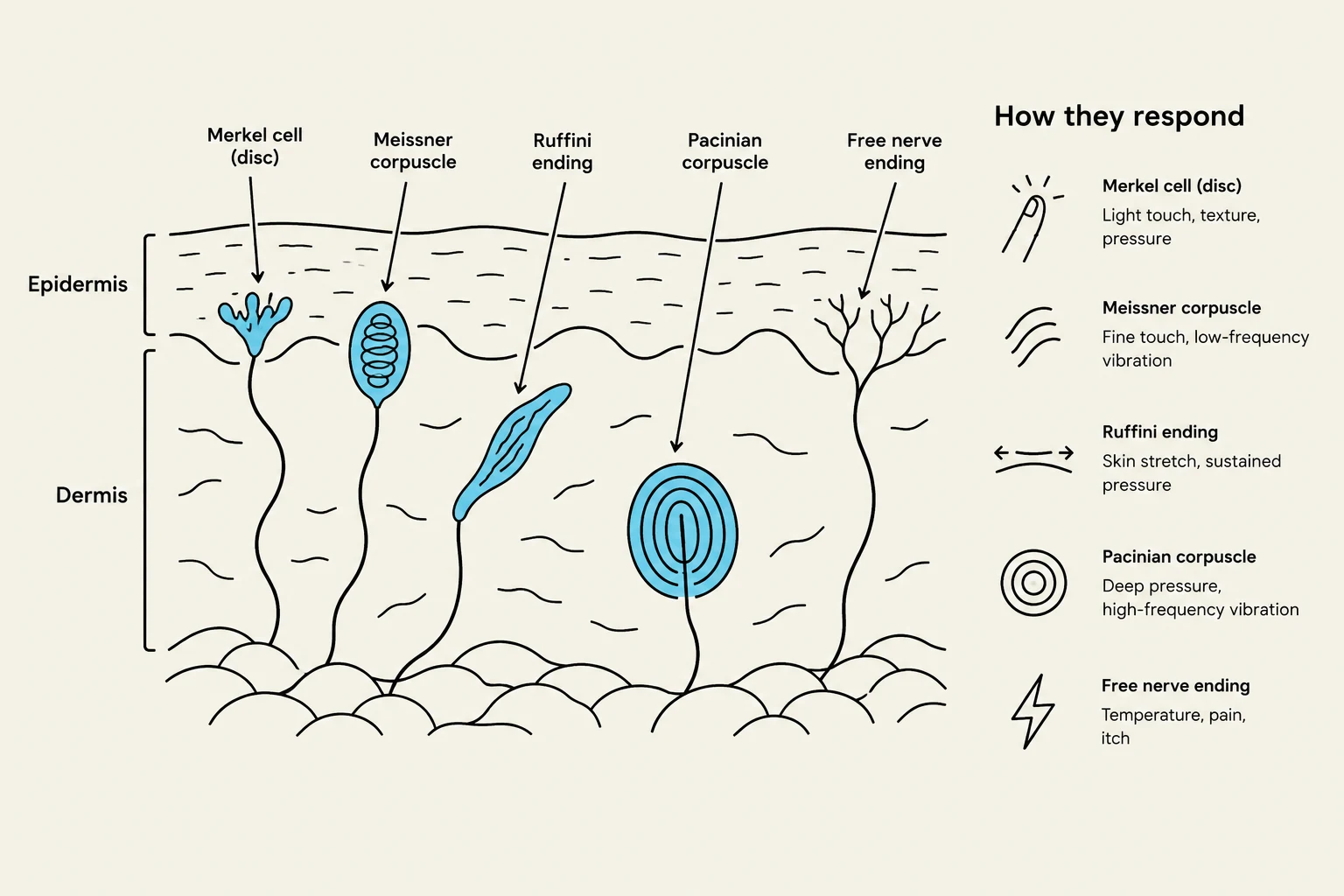
The skin acts as a massive sensory organ by housing various receptors that detect different stimuli. These include:
Tactile sensations: Touch, pressure, vibration, and tickle.
Thermal sensations: The ability to distinguish between warm and cool.
Pain: Detection of potentially damaging stimuli.
This diagram illustrates various mechanoreceptors in the skin, including Merkel cells, Meissner corpuscles, Ruffini endings, Pacinian corpuscles, and free nerve endings, along with their respective functions in sensing touch, pressure, vibration, temperature, and pain.
Clinical Considerations: Aging and Injury
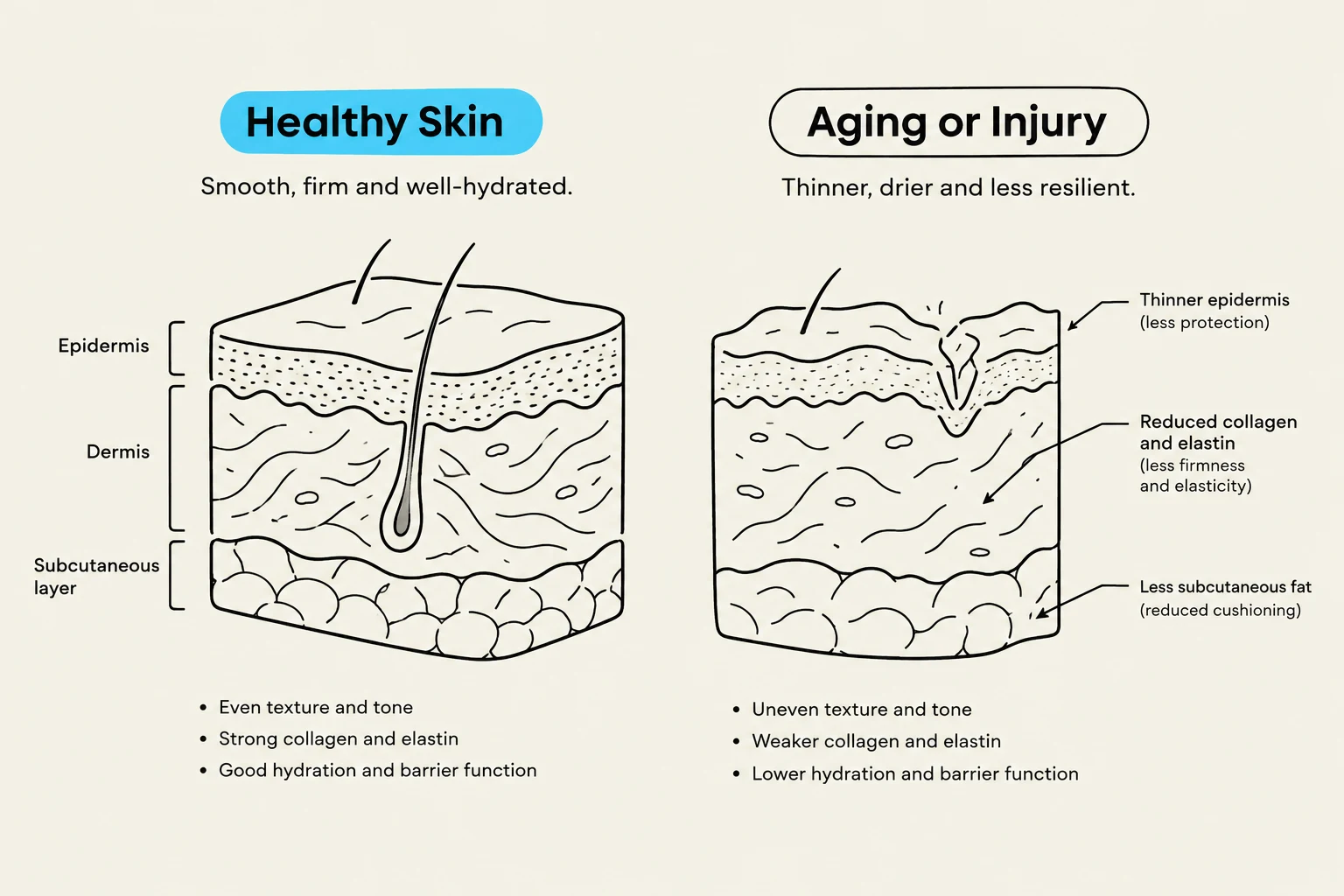
As the body ages, the integumentary system undergoes noticeable changes. These include the development of wrinkles, decreased sweat production, and a loss of subcutaneous fat, which leads to thinner skin. Additionally, a decrease in functional melanocytes can result in gray hair and atypical pigmentation.
The system is also susceptible to various injuries:
Skin Cancer: Often caused by excessive UV exposure, the three major types are basal cell carcinoma, squamous cell carcinoma, and malignant melanoma.
Burns: Tissue damage caused by heat, electricity, or chemicals that denature proteins.
Pressure Ulcers: Also known as "bed sores," these occur when a deficiency in blood flow causes the shedding of epithelium.
Diagram illustrating the structural differences between healthy skin and skin affected by aging or injury, highlighting changes in the epidermis, dermis, and subcutaneous layers.
Keep reading for free.
Create an account to finish this reading and make it part of your study routine.