An explanation of the body's inflammatory response, detailing the chemical alarm system, physiological changes like redness and swelling, and the cellular combatants involved in defending against pathogens.
Imagine you are walking through a garden and accidentally step on a thorn. Within minutes, the area around the puncture begins to feel warm, looks red, starts to swell, and becomes painful. While these sensations are uncomfortable, they are actually signs that your body has successfully identified a breach in its defenses and has mobilized its troops.
This process is known as the inflammatory response. It serves as the initial "field of battle" for the immune system, occurring when pathogens like bacteria or viruses bypass your first line of defense—the skin or mucous membranes. Because the response reacts similarly regardless of whether the intruder is a splinter or a virus, it is categorized as a nonspecific immune reaction.
The Chemical Alarm System
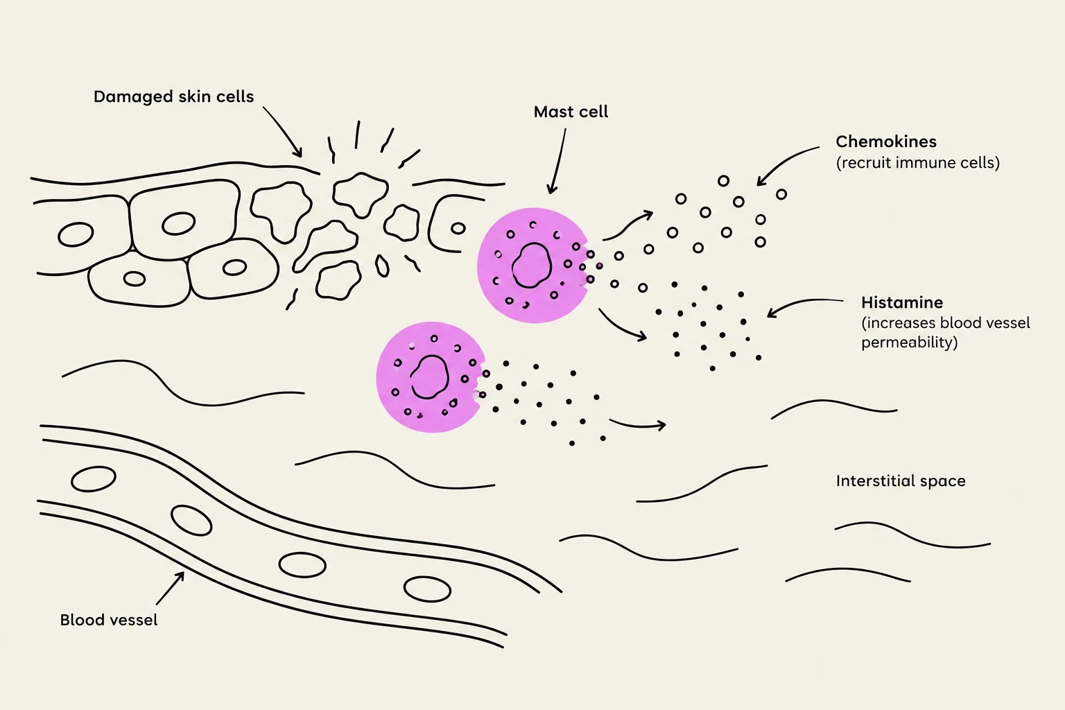
The battle begins the moment damage occurs. When cells are punctured or destroyed, they release chemical messengers known as chemokines. The name is derived from "chemo" (chemical) and "kine" (movement), reflecting their role as signaling proteins that direct immune cells to move toward the site of injury.
Simultaneously, specialized cells called mast cells are activated. These cells can be triggered by direct contact with a pathogen, by the chemokines released from damaged tissue, or even by molecules released by the invading bacteria themselves. Once activated, mast cells release a potent chemical called histamine.
Study Tip: Think of chemokines as the "GPS coordinates" that tell immune cells where to go, while histamine is the "alarm bell" that changes the local environment to allow those cells to arrive.
This diagram illustrates the inflammatory response, showing damaged skin cells, mast cells releasing histamine and chemokines, and their effects on blood vessels and the interstitial space.
Physiological Changes: Redness, Heat, and Swelling
Histamine is one of the primary drivers of the physical symptoms we associate with inflammation. It acts directly on the capillaries—the tiny blood vessels that deliver nutrients and oxygen to tissues—to facilitate two major changes:
Vasodilation: Histamine causes the endothelial cells (the cells that form the lining of the capillary walls) to signal the vessel to widen. This widening, or vasodilation, increases blood flow to the injured area, which results in the visible redness and the sensation of heat.
Increased Permeability: As the endothelial cells respond to histamine, they begin to separate slightly from one another. This makes the capillary walls more porous, allowing substances to leak out of the bloodstream and into the interstitial fluid (the fluid that bathes the cells).
As this fluid, along with proteins and immune cells, leaks out of the vessels, it creates a buildup called exudate. This accumulation of fluid in the tissue is what causes the characteristic swelling and contributes to the sensation of pain.
This diagram illustrates the process of vasodilation, where histamine causes endothelial cells to separate, increasing permeability and allowing fluid and proteins to leak into surrounding tissue, ultimately leading to increased blood flow.
The Cellular Combatants
Once the "gates" of the capillaries are opened, various immune cells rush to the field of battle. These cells can be divided into nonspecific responders and those that prepare the way for a specific attack.
The First Responders: Phagocytes
Phagocytes are specialized cells that protect the body by ingesting (eating) harmful foreign particles.
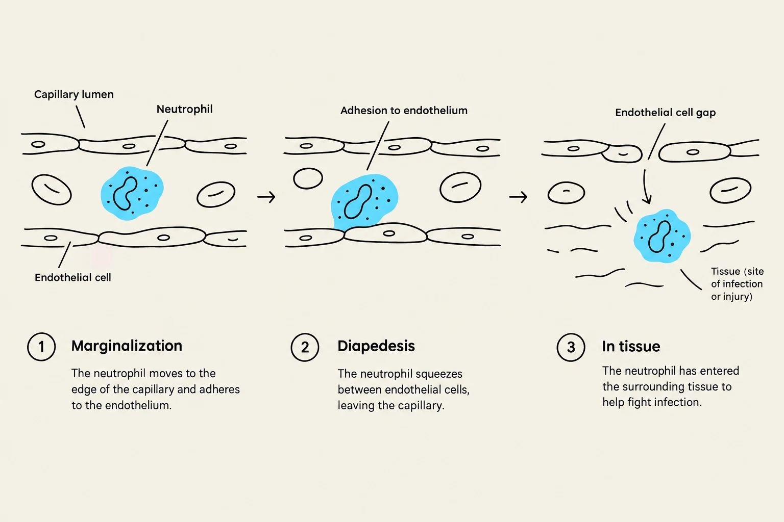
Neutrophils: These are the most abundant type of phagocyte in the body. They follow the chemokine gradient to the site of infection. To enter the tissue, they undergo a process called marginalization, where they roll along the inner walls of the capillary. They then perform diapedesis (also known as extravasation), which is the process of squeezing through the gaps between endothelial cells to reach the infection site.
Dendritic Cells: These phagocytes reside near external interfaces like the skin. They are highly efficient at ingesting pathogens and "presenting" them on their surfaces to other immune cells, acting as a bridge between the nonspecific and specific immune systems.
The Complement System: This is a group of plasma proteins that circulate in the blood in an inactive state. During inflammation, they become activated and can kill pathogens in a nonspecific manner, providing an extra layer of defense.
The Specialized Forces: B-cells and T-cells
While the initial response is nonspecific, the inflammatory environment also allows for the arrival of the adaptive immune system. B-cells and T-cells migrate to the site via the same processes of marginalization and diapedesis. Once they arrive and encounter the pathogens presented by dendritic cells, they can initiate a highly targeted, specific immune response tailored to that exact intruder.
This diagram illustrates the process of diapedesis, where a neutrophil moves from the capillary lumen to the surrounding tissue to combat infection or injury. It shows three stages: marginalization, adhesion to the endothelium, and passage through the endothelial cell gap.
Summary of the Inflammatory Bridge
The inflammatory response is much more than just a set of symptoms; it is a highly coordinated biological event. By altering the local physiology through vasodilation and increased permeability, the body creates a pathway for both the immediate, nonspecific defenders (like neutrophils and the complement system) and the long-term, specific defenders (like B-cells and T-cells) to meet the threat.
Exam Concept Check:
Redness/Heat→ caused by Vasodilation (increased blood flow).
Swelling→ caused by Increased Permeability (exudate entering interstitial fluid).
Pain→ caused by chemical mediators and tissue pressure from swelling.
Keep reading for free.
Create an account to finish this reading and make it part of your study routine.