This content provides a comprehensive overview of the cardiovascular system, detailing its three main circuits (pulmonary, systemic, and lymphatic), the anatomy and function of the heart as a central pump, and the hemodynamic forces that drive blood circulation.
The cardiovascular system is a sophisticated, life-sustaining network of interconnected circuits designed to transport essential substances throughout the body. Its primary mission is to deliver oxygen and nutrients to tissues while simultaneously removing metabolic waste products, such as carbon dioxide, for disposal. By maintaining this constant flow, the system ensures that every cell in the body receives the resources necessary for survival and homeostatic balance.
The Three Interconnected Circuits
The cardiovascular system does not function as a single loop, but rather as three distinct yet integrated circuits that work in tandem to manage blood flow and fluid balance.
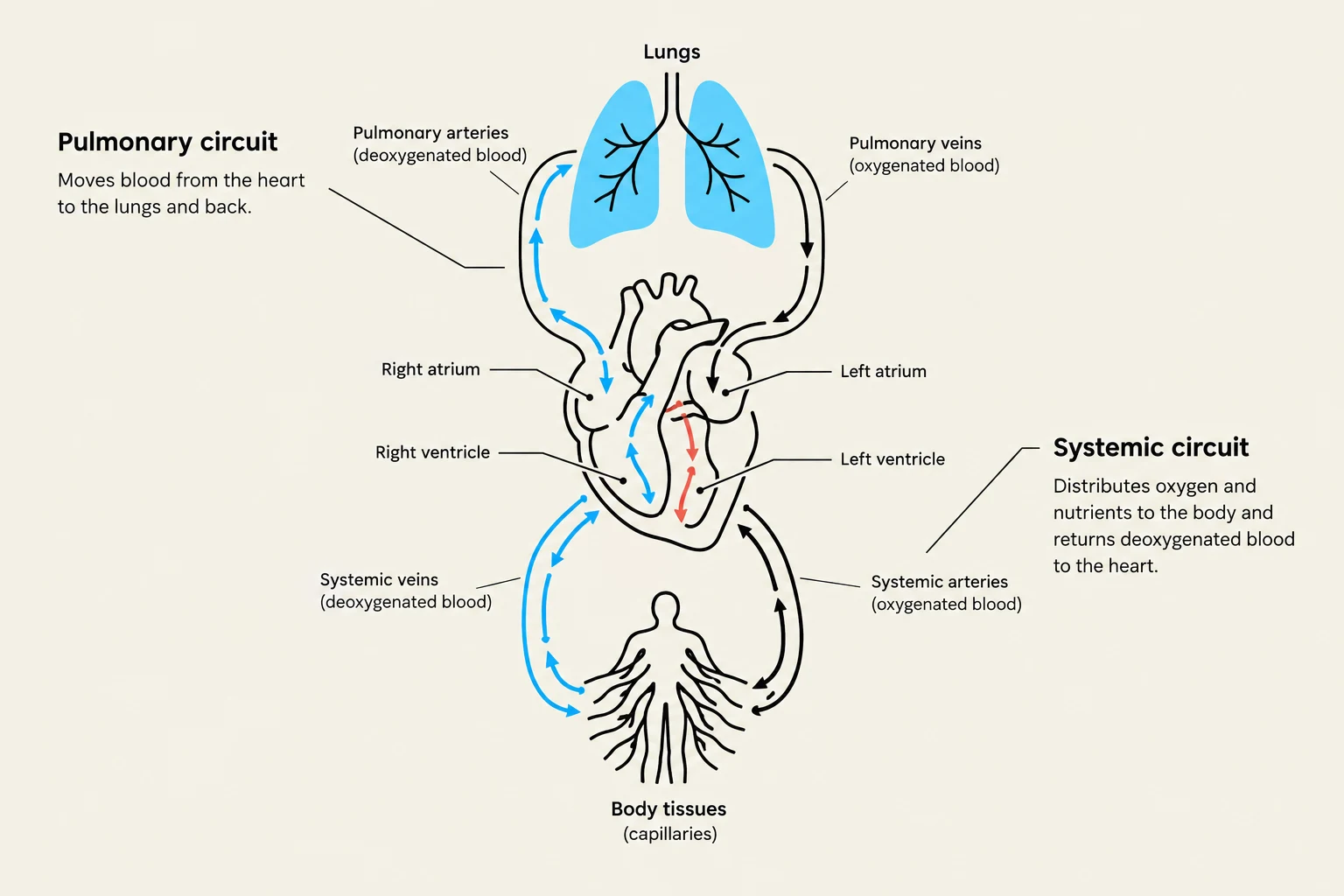
The Pulmonary Circuit: This circuit is responsible for moving blood from the right side of the heart to the lungs. In the lungs, the blood undergoes gas exchange: it unloads carbon dioxide (CO2) and loads oxygen (O2).
The Systemic Circuit: Once oxygenated, the blood returns to the left side of the heart and is pumped out through the systemic circuit to the rest of the body. Here, it delivers O2 to organs and tissues and picks up metabolic waste.
The Lymphatic Circuit: While often studied separately, the lymphatic circuit is a vital component of the broader circulatory system. It collects interstitial fluid (the fluid surrounding cells) from all over the body and returns it to the venous circulation, while also playing a major role in the immune system.
Diagram illustrating the pulmonary and systemic circuits of blood circulation in the human body, showing the heart, lungs, and body tissues.
Anatomy of the Heart: The Central Pump
At the center of this entire operation is the heart, an extraordinarily efficient and reliable pump that weighs less than a pound in humans. Despite its small size, it is capable of beating more than 3 billion times over an average human lifetime.
Chambers and Structure
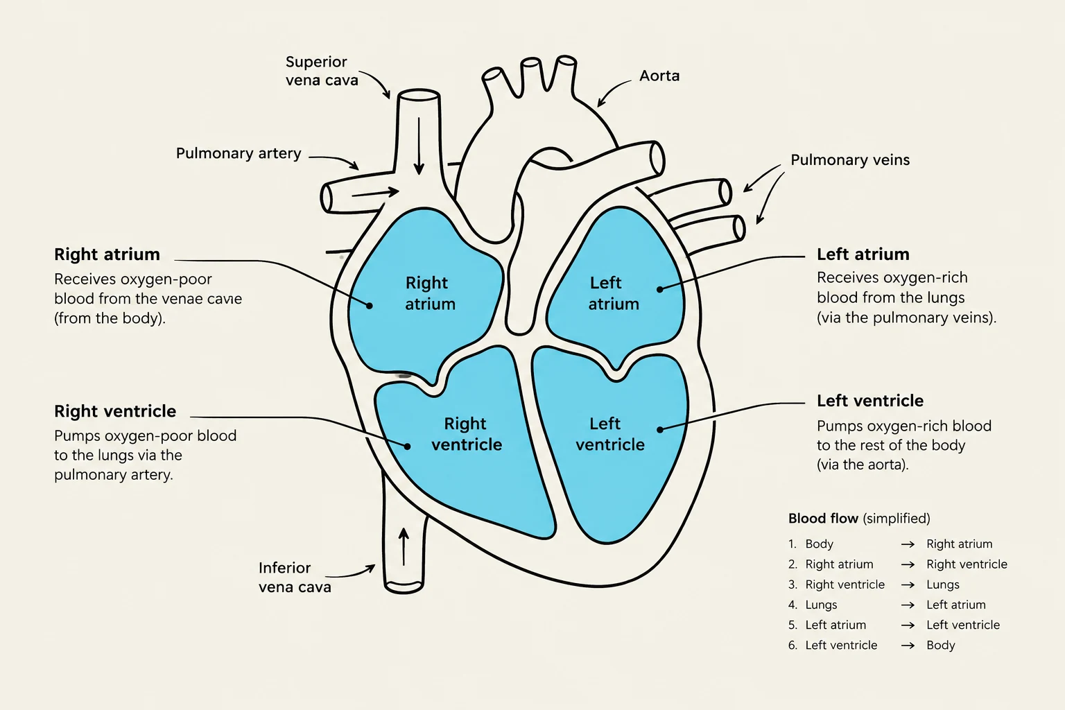
The heart is divided into four distinct chambers:
Atria: The two upper chambers (right and left atria) act as receiving rooms for venous blood.
Ventricles: The two lower chambers (right and left ventricles) are the primary powerhouses that push blood out into the arteries. The right ventricle specifically drives blood toward the lungs, while the left ventricle drives blood through the entire body.
These chambers are separated by a dense connective tissue known as the fibrous skeleton. This skeleton forms rings called the annuli fibrosus around the heart valves, providing structural support for the valve flaps.
The Role of Valves
To ensure that blood flows in only one direction, the heart utilizes several types of valves:
Atrioventricular (AV) Valves: Located between the atria and ventricles, these prevent backflow into the atria during ventricular contraction. The right side features the tricuspid valve (three flaps), while the left side features the bicuspid (or mitral) valve (two flaps).
Semilunar Valves: Located at the base of the large arteries leaving the heart, these allow blood to enter the pulmonary and systemic circuits when the ventricles contract, but prevent it from leaking back into the ventricles when they relax.
Diagram illustrating the four chambers of the heart (right atrium, right ventricle, left atrium, left ventricle) and the flow of blood through them, including major blood vessels like the aorta, pulmonary artery, pulmonary veins, superior vena cava, and inferior vena cava. A simplified blood flow sequence is also provided.
The Pathway of Blood Flow
Understanding the sequence of blood movement is essential for grasping how the circuits interact. The process follows a strict, unidirectional path:
Entry: Deoxygenated venous blood enters the right atrium via the superior and inferior venae cavae.
To the Right Ventricle: Blood passes through the tricuspid valve into the right ventricle.
To the Lungs: The right ventricle contracts, forcing the pulmonary valve open and sending blood through the pulmonary arteries to the lungs.
Return from Lungs: Oxygenated blood returns from the lungs via the pulmonary veins to the left atrium.
To the Left Ventricle: Blood flows through the bicuspid (mitral) valve into the left ventricle.
Systemic Distribution: The left ventricle contracts, forcing the aortic valve open and pushing blood into the aorta, where it is distributed to every organ in the body.
Hemodynamics: Driving Forces of Circulation
While the contraction of the ventricles provides the primary power for circulation, the heart does not work in isolation. Several auxiliary factors contribute to the total cardiac output (the volume of blood pumped by the heart per unit of time).
Venous Return
A significant portion of ventricular filling is actually driven by the pressure of returning blood rather than the contraction of the atria. At rest, atrial contraction only accounts for approximately 25% of ventricular filling; the remaining 75% is driven by the continuous, low-pressure flow of blood from the veins.
Because the veins hold between 60% and 70% of the body's total blood volume, the rate of venous return has a massive impact on cardiac output.
Clinical Note: Changes in venous return can be dramatic. Physical exercise, muscle contractions, or even changes in G-force (such as flying in a jet) can alter how much blood returns to the heart, directly affecting the heart's ability to pump.
Arterial Elasticity
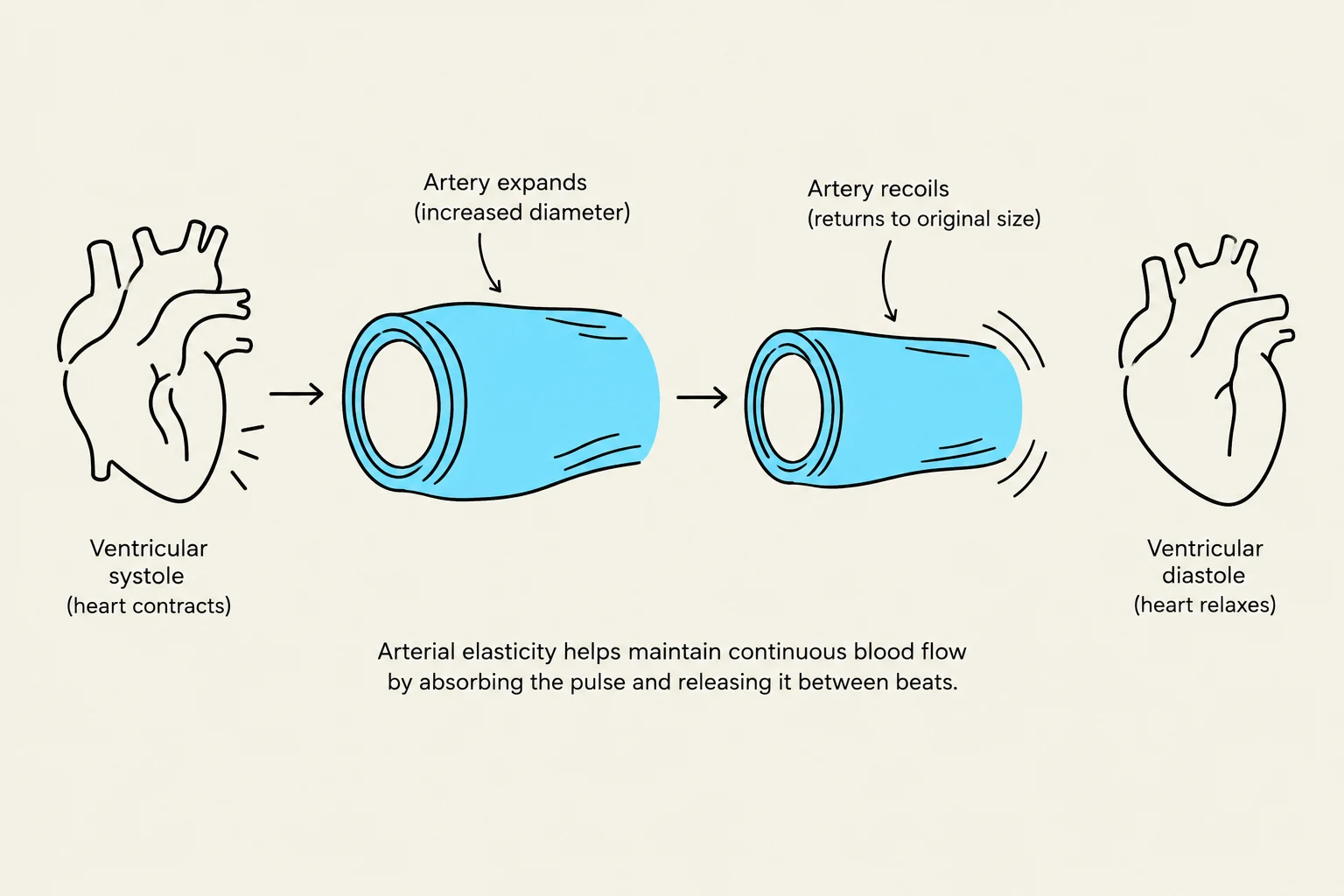
The major arteries also act as a secondary driving force through their inherent elasticity. As the ventricles contract, the walls of these large vessels expand like balloons to accommodate the surge of blood. When the ventricles relax, the elastic walls "spring back," which helps continue pushing blood through the circulation and "dampens" extreme pressure fluctuations that might otherwise damage smaller vessels (arterioles).
This diagram illustrates how arterial elasticity helps maintain continuous blood flow by absorbing the pulse during ventricular systole (heart contraction) and releasing it during ventricular diastole (heart relaxation).
Summary Table: Heart Valve Comparison
Valve Type
Location
Number of Flaps
Function
Tricuspid (AV)
Right Atrium → Right Ventricle
3
Prevents backflow to Right Atrium
Bicuspid/Mitral (AV)
Left Atrium → Left Ventricle
2
Prevents backflow to Left Atrium
Pulmonary (Semilunar)
Right Ventricle → Pulmonary Artery
N/A
Prevents backflow to Right Ventricle
Aortic (Semilunar)
Left Ventricle → Aorta
N/A
Prevents backflow to Left Ventricle
Keep reading for free.
Create an account to finish this reading and make it part of your study routine.