


Understanding the complexities of dental anatomy is fundamental to providing effective clinical care, whether you are performing periodontal probing, placing a restoration, or charting a patient's progress. To communicate effectively within a dental team, clinicians must master a specific vocabulary that describes the location, type, and physical structure of teeth. This article provides a comprehensive overview of dentition periods, tooth organization, and the intricate anatomy of the tooth itself.
Dentition Periods and Tooth Types
A person experiences two distinct dentitions throughout their lifetime: the primary (deciduous) dentition and the permanent dentition. While we often refer to primary teeth as "baby teeth," they are not merely temporary placeholders; they are critical for guiding eruption, maintaining space for permanent teeth, and supporting speech and facial development.
The transition between these two dentitions occurs in three distinct periods:
- Primary Period: Only primary teeth are present (approx. 6 months to 6 years).
- Mixed Period: Both primary and permanent teeth are present (approx. 6 to 12 years).
- Permanent Period: Usually only permanent teeth are present (starting after age 12).
The types of teeth present change significantly between these stages. Primary dentition consists of 20 teeth: 8 incisors, 4 canines, and 8 molars. Notably, premolar teeth are absent in the primary dentition; they only appear in the permanent dentition. Permanent dentition consists of 32 teeth, including incisors, canines, premolars, and molars.
Exam Tip: Remember that permanent molars are "nonsuccedaneous." This means they do not replace any primary teeth; instead, they erupt distal to the primary dentition.
Organizing the Oral Cavity: Quadrants, Sextants, and Naming
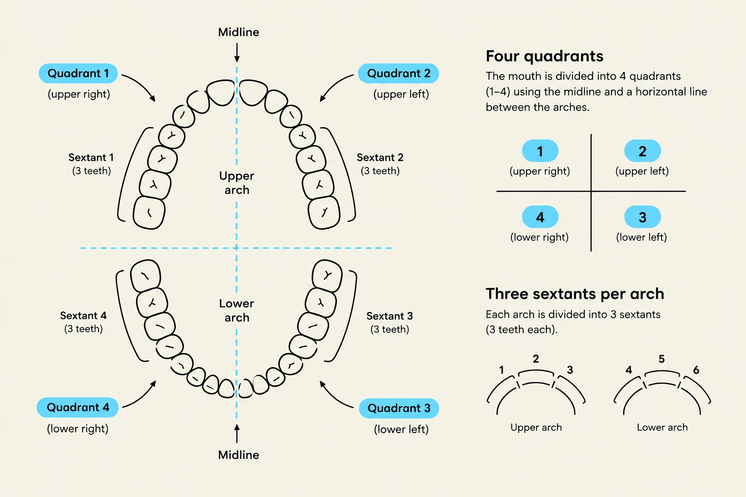
To ensure precision in charting, the oral cavity is organized into specific regions. Each dental arch (maxillary or mandibular) is divided by the midline into two quadrants.
Keep reading for free.
Create an account to finish this reading and make it part of your study routine.
Continue reading freeAlready have an account? Log in
Updated