Coagulation disorders, also known as coagulopathies, are complex clinical conditions arising from significant alterations in the body's ability to manage blood clotting. These disorders typically stem from abnormalities in either platelets (the cellular components of clotting) or clotting factors (the proteins that facilitate the coagulation cascade). The underlying causes can vary widely, ranging from autoimmune reactions and extensive blood loss to the formation of microemboli. Understanding the distinction between these disorders is critical, as some lead to excessive bleeding, while others paradoxically increase the risk of life-threatening clots.
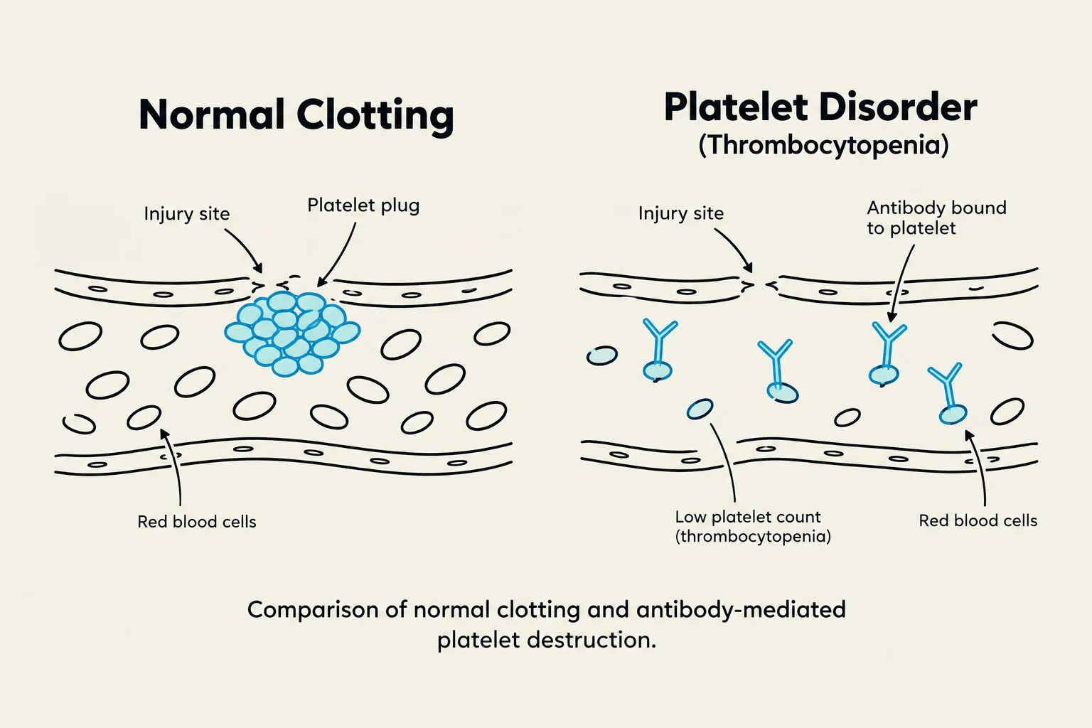
Comparison of normal clotting and platelet disorder (thrombocytopenia), illustrating the formation of a platelet plug at an injury site in normal clotting versus antibody-mediated platelet destruction in thrombocytopenia.
Autoimmune Platelet Disorders: ITP and TTP
Two primary autoimmune conditions affect platelet function and quantity: Idiopathic Thrombocytopenic Purpura (ITP) and Thrombotic Thrombocytopenic Purpura (TTP). While they share similar names, their pathophysiology and clinical risks are distinct.
Idiopathic Thrombocytopenic Purpura (ITP)
ITP is an autoimmune disorder where the body produces antiplatelet antibodies. These antibodies target the platelets, significantly decreasing their normal lifespan. As a result, the patient experiences a low platelet count, which can lead to severe hemorrhage.
Assessment and Diagnosis for ITP:
Laboratory Findings: Decreased hemoglobin (Hgb) and decreased platelet counts.
Screening: Clinicians often test for underlying triggers such as HIV, Hepatitis C, and H. pylori.
Bone Marrow Aspiration: This procedure is used to assess megakaryocytes, which are the precursor cells responsible for making platelets.
Management of ITP:
Treatment focuses on suppressing the immune response or increasing platelet production. Common interventions include:
Corticosteroids: Used to reduce the autoimmune response, though typically not for long-term use.
Thrombopoietin receptor agonists: Medications like Romiplostim and Eltromopag to stimulate platelet production.
Splenectomy: Considered if the patient does not respond to medical management.
Clinical Note: In ITP, platelet transfusions are generally not helpful because the patient's own antibodies will simply bind to and destroy the transfused platelets.
Thrombotic Thrombocytopenic Purpura (TTP)
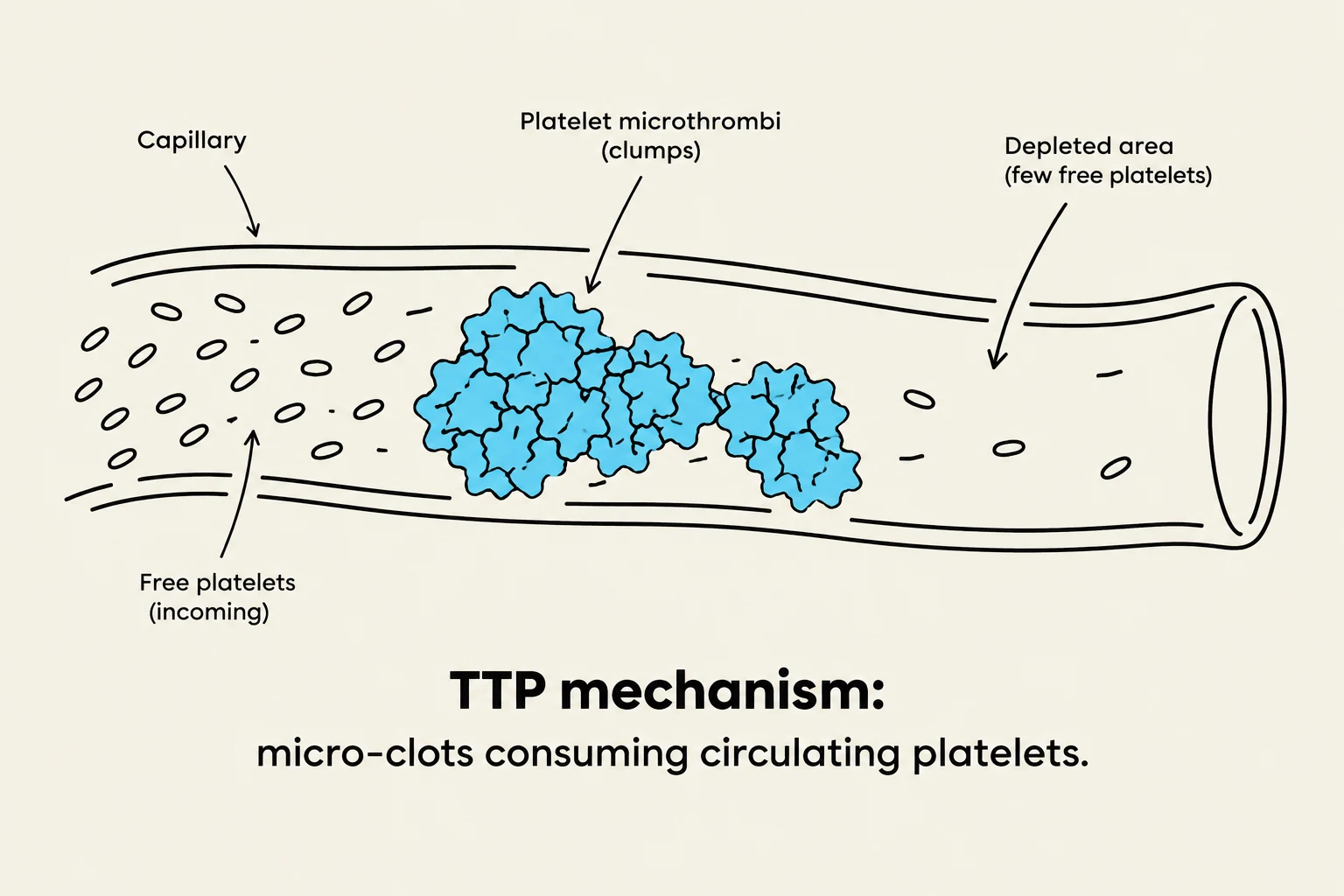
TTP is also driven by an autoimmune reaction, but instead of just destroying platelets, it causes abnormal platelet clumping and inappropriate clotting. This "clumping" uses up the available platelets, resulting in an insufficient quantity of platelets circulating in the bloodstream. This creates a dual danger: the lack of platelets causes bleeding, while the micro-clots can lead to serious complications like Renal Failure (RF), Myocardial Infarction (MI), or Cerebrovascular Accident (CVA).
Management of TTP:
Plasma exchange: A critical intervention to remove the offending antibodies.
Corticosteroids and Immunosuppressants: To manage the autoimmune trigger.
Antiplatelet medications: Such as Aspirin.
Warning: Unlike ITP, platelet transfusions in TTP patients can be dangerous, as they may increase the risk of a fatal thrombotic event due to the abnormal clumping mechanism.
Heparin-Induced Thrombocytopenia (HIT)
Heparin-Induced Thrombocytopenia (HIT) is an immunity-mediated clotting disorder that occurs as a reaction to the administration of heparin. Despite the name suggesting a low platelet count (thrombocytopenia), the primary danger is the increased risk of clotting.
When a patient develops HIT, the standard treatment is to discontinue heparin and switch to alternative anticoagulants that utilize a direct thrombin inhibitor, such as Argatroban or Bivalirudin.
Diagram illustrating the mechanism of TTP, where micro-clots consume circulating platelets within a capillary.
Disseminated Intravascular Coagulation (DIC)
Disseminated Intravascular Coagulation (DIC) is perhaps the most complex coagulation disorder. It is not a disease in itself, but rather a secondary complication triggered by an "insult" to the body, such as sepsis, trauma, cancer, obstetric complications (like abruptio placentae), or cardiopulmonary arrest.
Pathophysiology: The Paradoxical Hemorrhage
This diagram illustrates the DIC paradox, showing simultaneous micro-clotting in capillaries leading to reduced blood flow and tissue ischemia, and a broken larger vessel wall where platelets and fibrinogen are exhausted, resulting in systemic bleeding.
In DIC, the body's clotting and anti-clotting mechanisms occur simultaneously. An initial insult causes the release of chemicals into the bloodstream that instigate widespread coagulation. This leads to the formation of numerous microemboli (small clots) throughout the vasculature, which can cause ischemia and organ failure.
However, this widespread clotting process rapidly depletes the body's supply of platelets and clotting factors. Once these resources are exhausted, the patient loses the ability to clot where needed, resulting in a paradoxical hemorrhage—where the patient is simultaneously clotting in the small vessels and bleeding from larger ones.
Diagnostics and Clinical Manifestations
Clinicians look for specific laboratory markers to identify DIC:
Decreased: Platelets and Fibrinogen.
Increased: PT, INR, aPTT, and D-Dimer (a marker of clot breakdown).
Clinical signs include:
Bleeding/oozing, petechiae, ecchymosis, and hematuria.
Systemic instability: Tachycardia, hypotension, respiratory distress, and potential organ failure.
Treatment Strategies for DIC
The priority in treating DIC is to find and treat the underlying cause (e.g., treating the infection causing sepsis). Supportive care is also vital, including oxygenation, fluid replacement, and cardiovascular support.
To correct the secondary effects of the depletion of clotting components, the following may be administered:
Platelets: To correct low levels and control bleeding.
Fresh Frozen Plasma (FFP): To replace various coagulation factors.
Cryoprecipitate: Specifically used to replace fibrinogen and factors V & VII.
Heparin: Used in certain contexts to inhibit microthrombi and improve organ perfusion.
Keep reading for free.
Create an account to finish this reading and make it part of your study routine.