This content provides an overview of common cardiovascular system disorders, including their pathophysiology, diagnostic approaches, and general treatment measures. It covers conditions such as angina pectoris, myocardial infarction, cardiac dysrhythmias, and cardiac arrest, along with diagnostic tools like ECG and blo
The cardiovascular system is a vital network responsible for transporting oxygen, nutrients, and hormones to tissues while removing metabolic waste. Because of its continuous workload, the system is highly susceptible to various disorders that serve as major causes of comorbidity and mortality. These disorders range from chronic conditions like hypertension to acute, life-threatening emergencies such as myocardial infarction and cardiac arrest. Understanding the pathophysiology, diagnostic markers, and emergency interventions is essential for clinical practice.
Diagnostic Approaches for Cardiovascular Function
To assess the health of the heart and blood vessels, clinicians utilize a variety of diagnostic tools. These tests help differentiate between structural defects, electrical abnormalities, and perfusion issues.
Electrocardiogram (ECG/EKG): This is the primary tool for the initial diagnosis and monitoring of arrhythmias, myocardial infarction, and pericarditis.
Cardiac Catheterization & Angiography: A catheter is passed through a large vein (usually the leg) toward the heart. Dye is injected to visualize the coronary arteries via an angiogram, allowing clinicians to see obstructions directly.
Blood Biomarkers: Testing for troponin is critical, as elevated levels indicate cardiac muscle damage. Other tests include electrolytes (Na & K), CK-MB, and lipid profiles.
Imaging and Other Tests: Chest X-rays show heart size and pulmonary congestion, while nuclear imaging (like SPECT) assesses myocardial perfusion and ischemia.
This diagram illustrates how an ECG and a troponin blood test can be used to assess heart health. An ECG checks heart rhythm, while a blood test measures troponin levels, which can indicate heart muscle damage. Elevated troponin suggests a heart attack, while normal levels make it less likely.
Angina Pectoris: Myocardial Oxygen Deficit
Angina pectoris is characterized by chest pain resulting from a deficit of oxygen to the myocardium. This occurs when the heart's demand for oxygen exceeds the supply, often due to atherosclerosis (narrowing of the arteries) or coronary artery spasms.
Angina is classified into three main types:
Classic (Exertional) Angina: Triggered by physical activity or stress when the heart works harder.
Variant Angina: Occurs at rest, often due to vasospasms.
Unstable Angina: Prolonged pain at rest that may indicate a recent onset of ischemia and often precedes a myocardial infarction.
Clinical Tip: If chest pain is not relieved by rest or the administration of nitroglycerin (a coronary vasodilator), it should be treated as a potential myocardial infarction and requires immediate emergency intervention.
Myocardial Infarction (MI)
A Myocardial Infarction (heart attack) involves the actual death of myocardial tissue due to prolonged ischemia. This is typically caused by a thrombus (clot) or a total obstruction of a coronary artery.
Classification and Pathophysiology
MIs are categorized based on ECG findings:
STEMI (ST-elevation MI): Indicates a complete or significant blockage.
NSTEMI (non-ST-elevation MI): Indicates partial or different types of ischemia.
When an artery is blocked, the affected tissue becomes necrotic. Because myocardial fibers do not regenerate, the dead tissue is eventually replaced by nonfunctional fibrous tissue. Rapid intervention is the highest priority; restoring blood supply within 20–30 minutes can prevent irreversible damage.
Complications
Serious complications of an MI include:
Cardiogenic Shock: Occurs when the left ventricle's pumping capability is markedly impaired.
Congestive Heart Failure (CHF): Resulting from reduced ventricular contractility and declining stroke volume.
Sudden Death: Can occur within the first hour of an MI in approximately 25% of cases.
Diagram illustrating the progression of coronary artery blockage from healthy to completely blocked, explaining the impact on blood flow and potential health risks.
Cardiac Dysrhythmias
Dysrhythmias are deviations from the normal heart rate or rhythm caused by damage to the conduction system, electrolyte imbalances, hypoxia, or drug toxicity.
Sinus and Atrial Abnormalities
Sinus Node Issues: The SA node is the heart's natural pacemaker. Abnormalities include bradycardia (heart rate <60 bpm) and tachycardia (heart rate >100 bpm). Sick sinus syndrome involves alternating between these two states.
Atrial Abnormalities:Atrial fibrillation is a common dysrhythmia where the atria fail to contract efficiently. This causes blood to pool in the atria, significantly increasing the risk of clot formation.
Atrioventricular (AV) Node and Ventricular Abnormalities
Heart Block: This refers to conduction delays at the AV node. It ranges from first-degree (a simple delay) to third-degree (a total block where the atria and ventricles beat independently, with ventricles firing at a slow rate of 30–45 bpm).
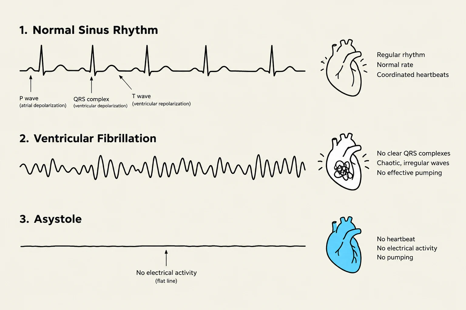
Ventricular Abnormalities: These are particularly dangerous. Ventricular tachycardia reduces cardiac output by decreasing filling time, while ventricular fibrillation is a life-threatening state where muscle fibers contract independently and rapidly. In fibrillation, the heart ceases to eject blood, leading to a total loss of cardiac output and severe hypoxia.
Cardiac Arrest (Asystole)
Cardiac arrest represents the complete cessation of all electrical and mechanical activity in the heart. On an ECG, this is identified as a "flatline" or asystole.
Because there is no cardiac output, the brain and heart are immediately deprived of oxygen, leading to a rapid loss of consciousness and cessation of respiration. Immediate resuscitation, often using Automated External Defibrillators (AEDs), is required to prevent brain death.
This illustration compares three different heart rhythms: Normal Sinus Rhythm, Ventricular Fibrillation, and Asystole, showing their corresponding electrocardiogram (ECG) readings and simplified heart diagrams.
Summary of General Treatment Measures
Management of cardiovascular disorders involves a combination of lifestyle modifications and pharmacological therapy:
Antihypertensives, diuretics, anticoagulants, and cholesterol-lowering drugs
Emergency
Oxygen, morphine, and thrombolytics (like tPA) for MI; defibrillation for life-threatening dysrhythmias
Exam Tip: Remember that in Congestive Heart Failure (CHF), "forward effects" refer to decreased cardiac output (fatigue, lethargy), while "backward effects" refer to congestion behind the failing ventricle (pulmonary edema in left-sided failure; systemic edema in right-sided failure).
Keep reading for free.
Create an account to finish this reading and make it part of your study routine.