Overview of Cardiac Disorders: From Coronary Artery Disease to Myocardial Infarction
This article explores the progression from coronary artery disease to acute myocardial infarction, detailing the clinical manifestations, diagnostic tools, and essential nursing interventions. It covers atherosclerosis, angina pectoris, acute coronary syndrome, signs and symptoms of AMI, diagnostic tools like ECG and c
Understanding cardiac disorders requires a grasp of how blood flow to the heart muscle is maintained. When the pathways providing oxygenated blood—the coronary arteries—become compromised, the heart muscle undergoes ischemia (lack of oxygen) and potentially infarction (cell death). This article explores the progression from coronary artery disease to acute myocardial infarction, detailing the clinical manifestations, diagnostic tools, and essential nursing interventions.
Coronary Artery Disease (CAD) and Atherosclerosis
Coronary Artery Disease (CAD) is a condition where the large coronary arteries become blocked. This blockage is primarily driven by atherosclerosis, a process characterized by inflammation, the accumulation of fatty deposits, and the subsequent hardening of the arterial walls.
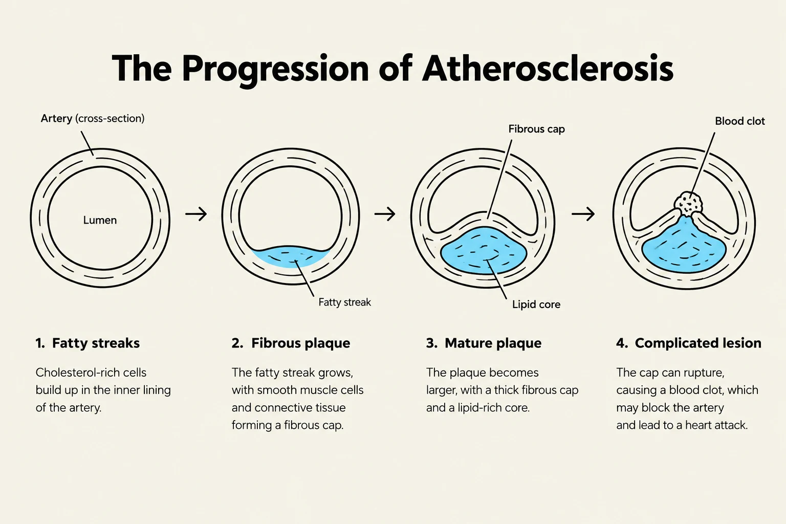
Atherosclerosis typically progresses through developmental stages:
Fatty streaks: The earliest visible sign of buildup.
Fibrous plaque: A more stable, hardened buildup.
Complicated lesions: Advanced stages that can lead to total occlusion.
Symptoms of CAD generally do not manifest until the blockage reaches more than 50% of the vessel's diameter.
The progression of atherosclerosis, illustrating the development of fatty streaks, fibrous plaques, mature plaques, and complicated lesions within an artery.
Risk Factors
Risk factors for CAD are categorized into two groups:
Non-Modifiable: Factors you cannot change, such as age, gender, ethnicity, and family history (which may be associated with increased C-reactive protein levels).
Modifiable: Factors that can be managed through lifestyle or medical intervention, including elevated serum lipids, hypertension, tobacco use, physical inactivity, obesity, and diabetes.
Angina Pectoris and Acute Coronary Syndrome
When the heart muscle experiences reversible ischemia due to blocked coronary arteries, the patient often experiences Angina Pectoris. Angina is categorized into three main types:
Stable (Chronic) Angina: This occurs most frequently during exercise or physical activity and typically subsides with rest.
Unstable Angina: A more dangerous form that occurs at rest or with minimal activity and is not relieved by nitroglycerin.
Variant (Prinzmetal) Angina: Caused by a coronary artery spasm rather than traditional atherosclerosis; it may not be associated with CAD.
When these conditions escalate, they fall under the umbrella of Acute Coronary Syndrome (ACS), which includes unstable angina and various forms of myocardial infarction.
Clinical Pearl: The 20-Minute Window
Myocardial tissue is highly sensitive to oxygen deprivation. Within 10 seconds of an occlusion, the tissue becomes hypoxic. The tissue remains viable for approximately 20 minutes before irreversible damage begins.
Acute Myocardial Infarction (AMI)
An Acute Myocardial Infarction (AMI) occurs when a coronary artery is completely occluded, leading to prolonged ischemia. If ischemia lasts longer than 20 minutes, it results in irreversible cell death. While collateral circulation can sometimes minimize the extent of the damage, the healing process often results in the formation of scar tissue, which leads to a permanent loss of cardiac function.
Signs and Symptoms
Patients presenting with AMI may exhibit "typical" symptoms, but clinicians must be alert for "atypical" presentations.
Typical Symptoms
Atypical Symptoms (High Risk)
Chest pain
Women
Shortness of breath (SOB)
People with diabetes
Diaphoresis (sweating)
The elderly
Cool, clammy skin
Nausea and vomiting
Anxiety or a "sense of doom"
Changes in vital signs
Diagnostic Tools: ECG and Cardiac Enzymes
To diagnose an AMI, healthcare providers rely on Electrocardiograms (ECG) and blood tests for cardiac enzymes.
1. ECG Findings
The ECG helps differentiate between types of myocardial injury:
Ischemia: Indicated by T-wave inversion.
Injury: Indicated by ST-segment elevation.
STEMI vs. NSTEMI: A STEMI (ST-Elevation Myocardial Infarction) shows ST-segment elevation on the ECG, whereas an NSTEMI (Non-ST-Elevation Myocardial Infarction) does not show ST elevation.
2. Cardiac Enzymes
When heart cells die, they release specific proteins into the bloodstream:
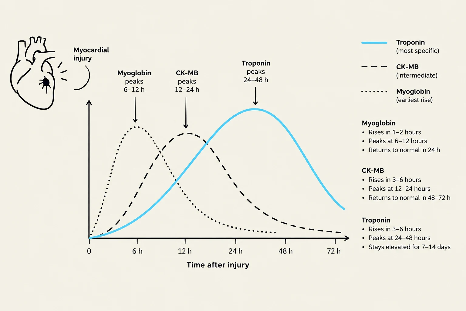
Troponin: This is a critical marker; levels rise approximately 3 hours after injury and peak at 12 hours.
CK & CK-MB: These rise 3–12 hours after injury and peak at 24 hours.
Myoglobin: Released very early (1–4 hours), but it is not specific to cardiac tissue.
This graph illustrates the typical time course of myoglobin, CK-MB, and troponin levels in the blood after a myocardial injury, such as a heart attack. Myoglobin rises earliest, followed by CK-MB, and then troponin, which is the most specific marker for myocardial injury and remains elevated for the longest period.
Medical Treatment and Nursing Care
Emergency Management: MONA
The primary goal in treating an AMI is to preserve myocardial tissue and prevent further cell death. A common mnemonic used for initial medical management is MONA:
Morphine
Oxygen
Nitroglycerin (Sublingual or IV)
Aspirin
Reperfusion and Surgical Interventions
For patients with a STEMI, emergent reperfusion is required. This can be achieved through Percutaneous Coronary Intervention (PCI) or Fibrinolytic therapy.
If a patient has extensive disease or if stent placement is unsuccessful, they may require a Coronary Artery Bypass Graft (CABG). Post-CABG care is intensive, often requiring ICU monitoring for 24–36 hours, management of chest tubes, and strict adherence to "sternal precautions".
Nursing Care and Complications
Nurses play a vital role in monitoring for life-threatening complications, including:
Dysrhythmias
Heart failure
Cardiogenic shock
Thromboembolism
Ventricular aneurysm or rupture
Standard nursing interventions include continuous ECG monitoring, vital sign assessment, maintaining bedrest to reduce cardiac workload, and managing pain.
Long-Term Management and Prevention
For patients surviving an acute event, the focus shifts to Cardiac Rehabilitation and lifestyle modification to prevent future events.
Medications: Long-term therapy often includes Statins, Beta-blockers, ACE inhibitors, and Antiplatelet medications (such as Aspirin or Clopidogrel).
Lifestyle: Emphasis is placed on nutritional therapy (low sodium, heart-healthy diets), smoking cessation, stress reduction, and supervised exercise.
Education: Patients must be educated on managing chronic conditions like hypertension and diabetes to reduce the overall burden on the heart.
Exam Tip: Stable Angina Management (The ABCDE Approach)
To remember the management of stable angina, use this mnemonic:
A – Aspirin, anti-angina agents, ACE inhibitors
B – Beta-blockers and blood pressure control
C – Cigarette smoking cessation and cholesterol management
D – Diet and diabetes management
E – Education and exercise
Keep reading for free.
Create an account to finish this reading and make it part of your study routine.