Anatomical notes detailing the osteology of the shoulder, glenohumeral joint movement, and muscle actions including innervation and distal attachments.
The shoulder is one of the most complex and mobile regions of the human body. Its functionality relies on a sophisticated interplay between various bony structures, joints, and a specialized group of muscles that provide both extreme range of motion and necessary stability. Understanding the shoulder requires looking beyond a single joint to the "shoulder complex," which integrates the glenohumeral joint and the scapulothoracic interface.
Osteology and the Shoulder Complex
The shoulder complex is not a single joint but a functional unit involving several key bony structures. The primary components include the humerus, the scapula, and the clavicle. A defining characteristic of the scapula is its orientation; it maintains a position approximately 30−40∘ anterior to the frontal plane.
The stability and movement of the shoulder are governed by the relationship between the glenohumeral (GH) joint and the scapulothoracic (ST) joint. A fundamental biomechanical principle used in clinical assessment is the movement ratio: for every $2''$ of glenohumeral joint movement, there is $1''$ of scapulothoracic movement.
The Glenohumeral Joint
The GH joint is a ball-and-socket articulation where the head of the humerus meets the glenoid cavity of the scapula. To facilitate smooth movement, the joint is enclosed by a joint capsule that contains synovial fluid. This capsule is reinforced by various ligaments that provide passive stabilization to the joint.
Scapulothoracic Articulation
The scapulothoracic joint is a functional rather than a true anatomical joint, consisting of the interface between the posterior rib cage (typically ribs 2-8) and the subscapularis/costal surface of the scapula. The scapula can undergo several distinct movements:
Elevation and Depression: Superior and inferior translation relative to the thoracic wall.
Adduction (Retraction): Medial translation toward the spine.
Abduction (Protraction): Lateral translation away from the spine.
Upward and Downward Rotation: Arcing of the glenoid fossa and inferior angle.
Exam Tip: Remember the 2:1 ratio. During abduction, the movement is shared between the GH joint and the scapula. If a patient lacks scapular upward rotation, they will struggle to achieve full overhead reach.
Rotator Cuff Mechanics
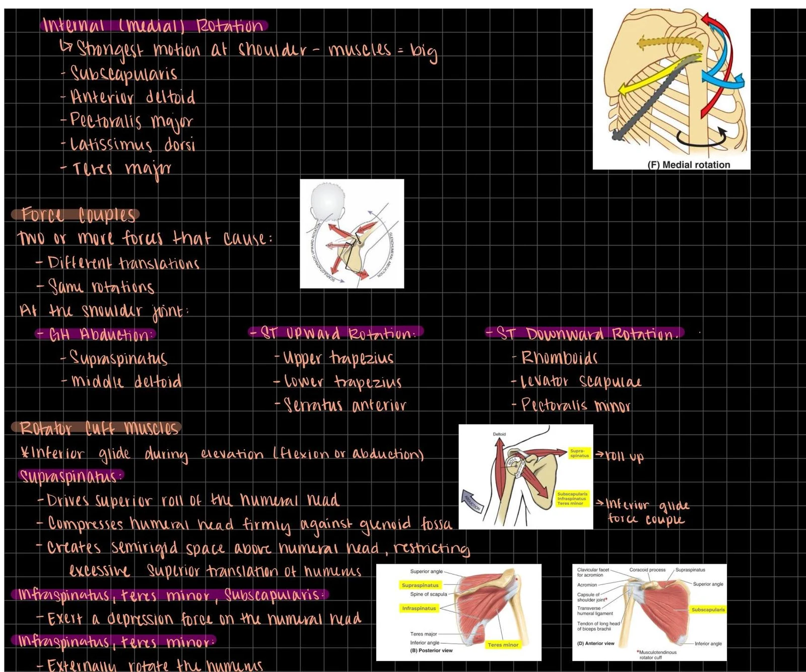
Because the glenoid cavity is relatively shallow compared to the large humeral head, the shoulder relies heavily on dynamic stability. This is provided by the rotator cuff, a group of muscles that work in tandem to stabilize the humeral head within the glenoid cavity.
The primary muscles of the rotator cuff include the supraspinatus, infraspinatus, teres minor, and subscapularis. Their collective action is to "compress" the humeral head into the socket, preventing dislocation during movement.
Diagrams illustrating shoulder joint anatomy and muscle actions, including internal rotation, force couples, and rotator cuff muscle functions.
Detailed Muscular Anatomy
The muscles of the shoulder can be categorized by their action on the GH joint versus the ST joint. Below is a detailed breakdown of the key muscles, their origins, distal attachments, innervation, and primary actions.
The Rotator Cuff Group
Muscle
Proximal Attachment
Distal Attachment
Innervation
Primary Action
Supraspinatus
Supraspinatus fossa of scapula
Superior fossa of greater tubercle
Suprascapular nerve
Initiates abduction; works with rotator cuff
Infraspinatus
Infraspinatus fossa of scapula
Middle fossa of greater tubercle
Suprascapular nerve
Lateral (external) rotation
Teres Minor
Lateral border of scapula
Inferior fossa of greater tubercle
Axillary nerve
Lateral (external) rotation
Subscapularis
Subscapular fossa (anterior scapula)
Lesser tubercle of humerus
Upper/Lower subscapular nerves
Medial (internal) rotation; stabilizes head
Additional Key Muscles
Deltoid
The deltoid is the most prominent muscle of the shoulder. It originates from the acromion and the lateral third of the clavicle, inserting on the lateral aspect of the humerus. It is innervated by the axillary nerve. Its actions are multi-faceted:
Anterior fibers: Flexion and medial rotation.
Middle fibers: Primary abductor of the arm.
Posterior fibers: Extension and lateral rotation.
Teres Major
Unlike the teres minor, the teres major is not part of the rotator cuff. It originates from the posterior lateral aspect of the inferior angle of the scapula and inserts on the intertubercular sulcus of the humerus. It is innervated by the lower subscapular nerve and assists in medial rotation and adduction.
Levator Scapulae
This muscle primarily influences scapular movement rather than humeral movement. It originates from the posterior processes of the upper cervical vertebrae and inserts on the superior angle of the scapula, innervated by the dorsal scapular nerve.
Clinical Summary of Innervation and Action
Understanding the neurovascular relationship is vital for diagnosing shoulder dysfunction. For example, injuries to the axillary nerve will directly impact both the deltoid and the teres minor.
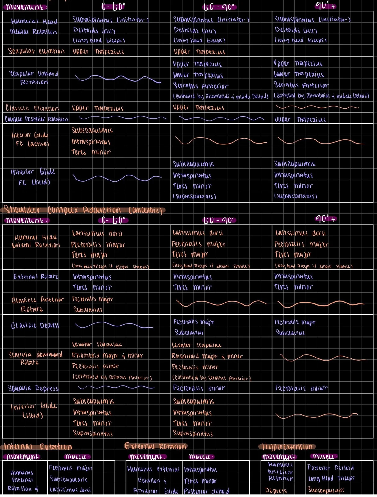
This chart illustrates the muscles involved in various shoulder and arm movements, categorized by the degree of rotation or angle.
When analyzing movement phases, clinicians often look at the degree of abduction:
Phase I (0-90°): Primarily involves scapular elevation and initial GH abduction.
Phase II (Above 90°): Scapular elevation stops, and the movement becomes primarily focused on scapular upward rotation and clavicular posterior rotation.
Clinical Note: If a patient can initiate abduction but cannot progress past 90°, the issue may lie in the scapulothoracic rhythm or the rotator cuff's ability to stabilize the humeral head during the transition to Phase II.
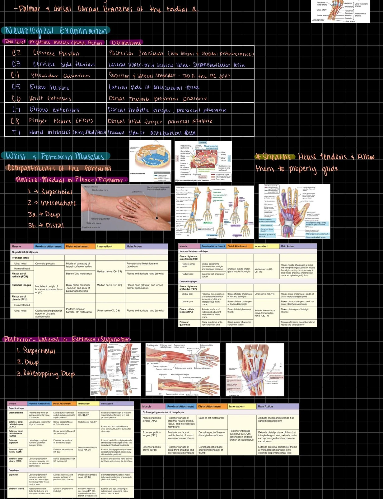
Anatomical illustrations and tables detailing the muscles of the forearm and wrist, their attachments, innervation, and actions, along with a neurological examination chart correlating spinal nerve roots to muscle actions and dermatomes.
Keep reading for free.
Create an account to finish this reading and make it part of your study routine.