Organization of the Vertebrate Nervous System and Circadian Rhythms
Explore the structure and function of the vertebrate nervous system, including the central and peripheral divisions, spinal cord, and reflex behaviors. Learn about homeostasis, the autonomic nervous system, and the molecular mechanisms of circadian rhythms regulated by the suprachiasmatic nucleus.
The vertebrate nervous system is a masterpiece of biological organization, ranging from the microscopic oscillations of individual genes to the complex, large-scale coordination of entire bodily systems. To understand how an organism maintains stability—such as regulating its internal clock or responding to a sudden threat—one must first understand the structural hierarchy of the nervous system and the specialized centers that govern homeostasis.
The Architecture of the Vertebrate Nervous System
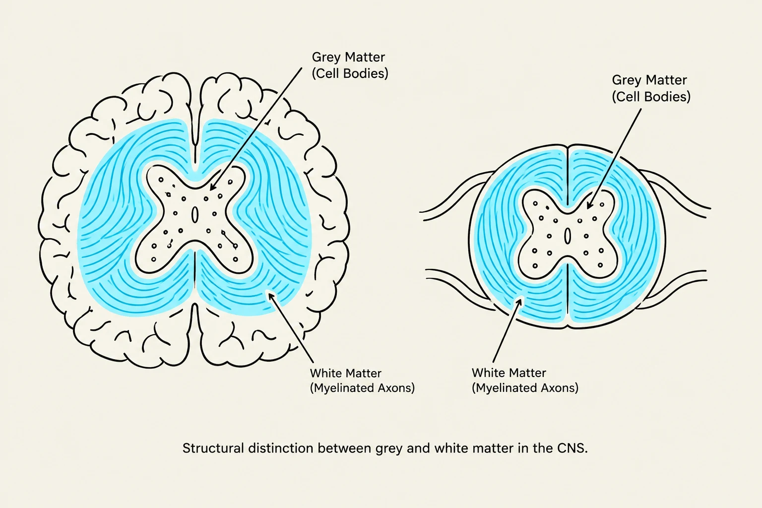
Structural distinction between grey and white matter in the CNS.
The vertebrate nervous system is divided into two primary anatomical divisions: the Central Nervous System (CNS) and the Peripheral Nervous System (PNS).
The Central Nervous System (CNS)
The CNS serves as the body's command and integration center, consisting of the brain and the spinal cord. Within the CNS, we distinguish between two types of tissue:
White matter: Composed primarily of myelinated axons, giving it a bright, shiny appearance.
Grey matter: Contains neuronal cell bodies, dendrites, and unmyelinated axons, appearing more dull.
The brain is further organized into functional regions. The hindbrain (including the medulla oblongata, pons, and cerebellum) manages vital functions like breathing and motor coordination. The forebrain (cerebrum) handles "higher" processes such as sensory perception and motor control. Deep within the forebrain lies the diencephalon, which contains the thalamus (a sensory relay station) and the hypothalamus.
The Peripheral Nervous System (PNS)
The PNS consists of all neurons located outside the CNS. It is organized into nerves (bundles of axons) and ganglia (clusters of cell bodies). A critical component of the PNS is the autonomic nervous system, which regulates involuntary physiological processes.
Exam Tip: Autonomic Divisions
Remember that the autonomic nervous system is split into two antagonistic branches: the Sympathetic system ("fight or flight") and the Parasympathetic system ("rest and digest").
Spinal Cord Organization and Reflex Behaviors
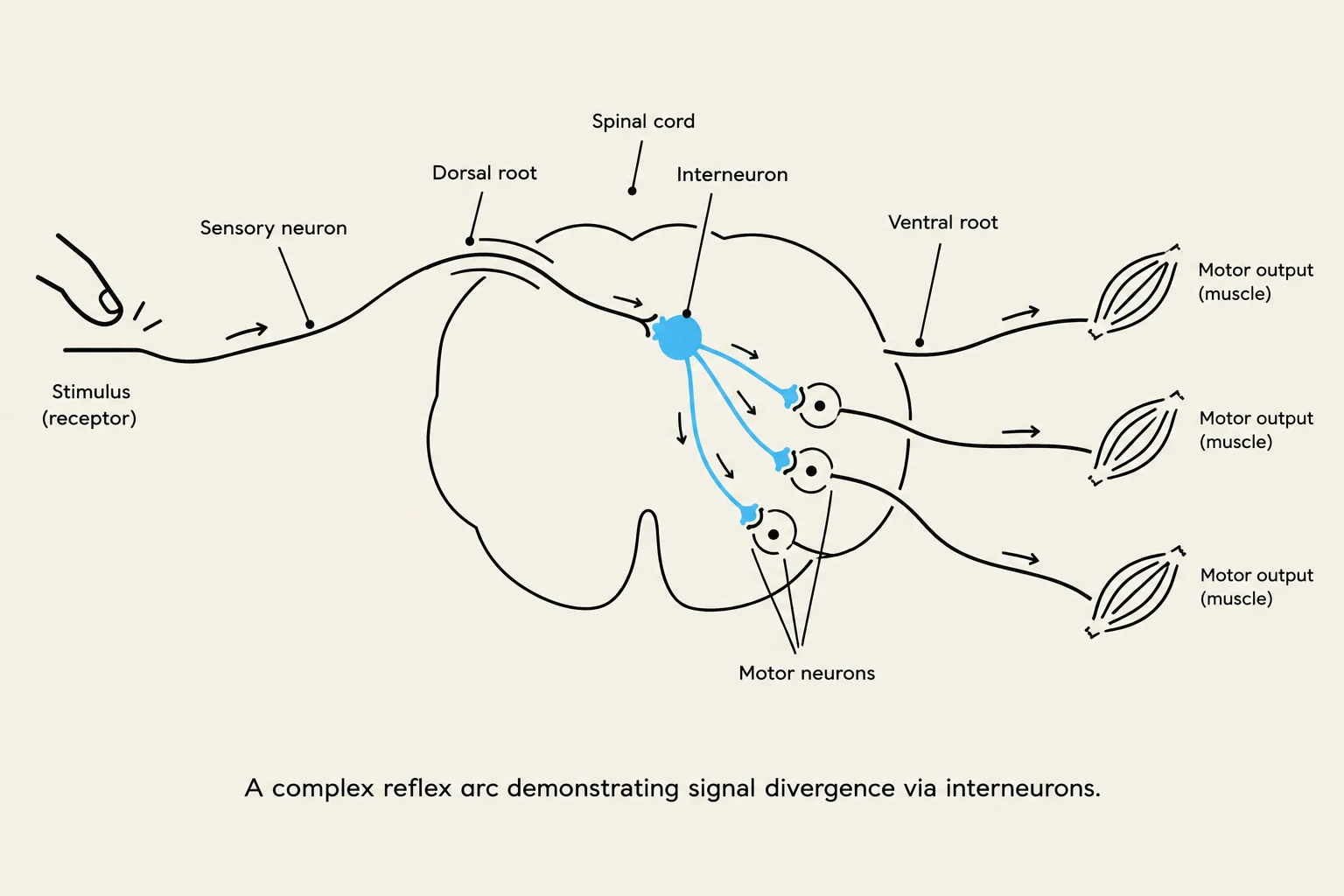
A complex reflex arc demonstrating signal divergence via interneurons.
The spinal cord acts as the primary pathway for information traveling between the brain and the body. It is organized such that sensory information enters through the dorsal root and motor commands exit through the ventral root.
One of the most fundamental functions of the spinal cord is the mediation of reflex behaviors. These are involuntary, rapid responses to stimuli.
Simple Reflex Arcs: A basic reflex, like the patellar (knee-jerk) reflex, may require only a single sensory neuron and a single motor neuron.
Complex Reflexes: More sophisticated responses, such as the withdrawal reflex, utilize interneurons and a process called divergence. In divergence, a single sensory neuron synapses onto an interneuron, which then spreads the signal to many different motor neurons to ensure a coordinated muscular response.
Homeostasis and the Autonomic Nervous System
To maintain a stable internal environment (homeostasis), the body relies on the balance between the sympathetic and parasympathetic systems.
Feature
Sympathetic System
Parasympathetic System
Primary Role
Stress / Physical Activity
Rest / Digestion
Nickname
"Fight or Flight"
"Rest and Digest"
Key Effects
↑ Heart rate, ↑ Breathing, ↑ Blood to muscles
↓ Heart rate, ↑ Digestion, energy maintenance
These systems often exhibit dual innervation, meaning they act antagonistically on the same organ. For example, the sympathetic system might increase heart rate while the parasympathetic system works to decrease it. The chemical messengers used in these pathways include Acetylcholine (ACh) and Norepinephrine.
Circadian Rhythms: The Biological Clock
While the autonomic nervous system manages immediate physiological shifts, the body also requires a mechanism to manage long-term, daily cycles. These are known as circadian rhythms—endogenous, approximately 24-hour cycles of behavior and physiology that persist even in the absence of external environmental cues.
The Suprachiasmatic Nucleus (SCN)
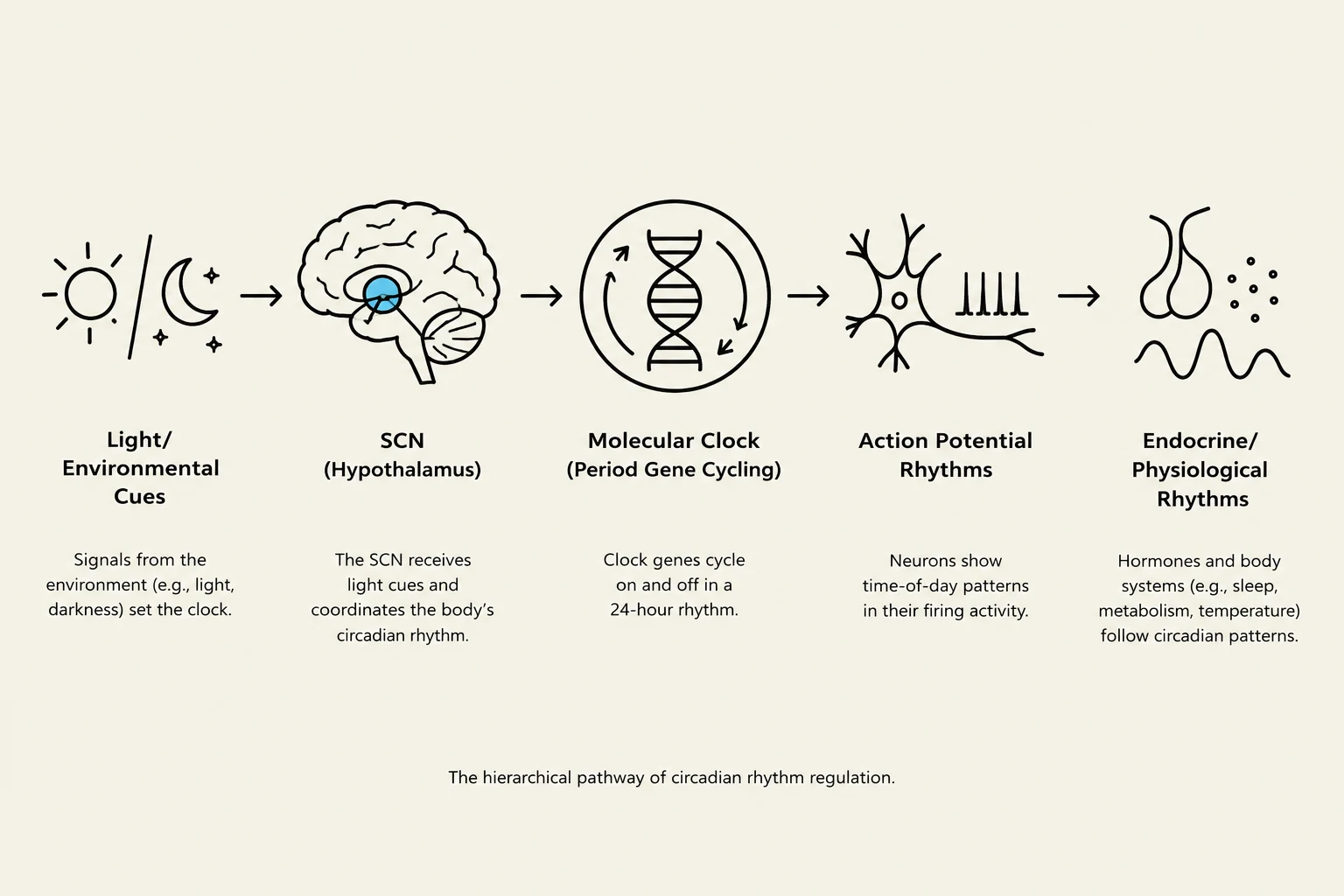
The hierarchical pathway of circadian rhythm regulation, starting from environmental cues like light and darkness, processed by the SCN (Hypothalamus), leading to molecular clock mechanisms, action potential rhythms in neurons, and finally endocrine/physiological rhythms.
The master control center for these rhythms is located in the hypothalamus. Specifically, a specialized structure called the suprachiasmatic nucleus (SCN) acts as the body's central pacemaker.
The SCN operates through a sophisticated input-output system:
Input Pathways: The SCN receives signals from environmental cues (such as light).
Output Pathways: The SCN sends signals to regulate various physiological rhythms, primarily by controlling the endocrine system.
The Molecular Mechanism: The Negative Feedback Loop
The "ticking" of the biological clock is not mechanical, but molecular. It is driven by a molecular negative feedback loop involving the rhythmic expression of specific genes, most notably the Period gene.
The process works as follows:
The expression of the Period gene fluctuates in a cycle.
This cycling of gene expression causes rhythmic changes in the action potential frequency of the neurons within the SCN.
This molecular oscillation ensures that the organism's internal biology remains synchronized with the Earth's light-dark cycle.
The discovery of these molecular mechanisms—explaining how gene expression drives the circadian rhythm—was so significant that it was recognized with the 2017 Nobel Prize in Physiology or Medicine.