


Skeletal muscles are not merely loose bundles of cells; they are highly organized biological machines. To function effectively, these muscles require a sophisticated structural framework that holds the tissue together, transmits force, and provides a pathway for essential life-support systems like blood and nerves. This organization is achieved through three distinct layers of connective tissue that wrap around different levels of the muscle structure.
The Macro Structure: Epimysium and Deep Fascia
At the largest scale, an entire skeletal muscle is encased in a protective outer layer known as the epimysium. This layer is composed of dense collagen fibers that serve a critical structural purpose: they separate the muscle from the surrounding organs and adjacent tissues.
The epimysium does not exist in isolation; it is continuous with the deep fascia, a dense layer of connective tissue that helps anchor the muscle to the rest of the body.
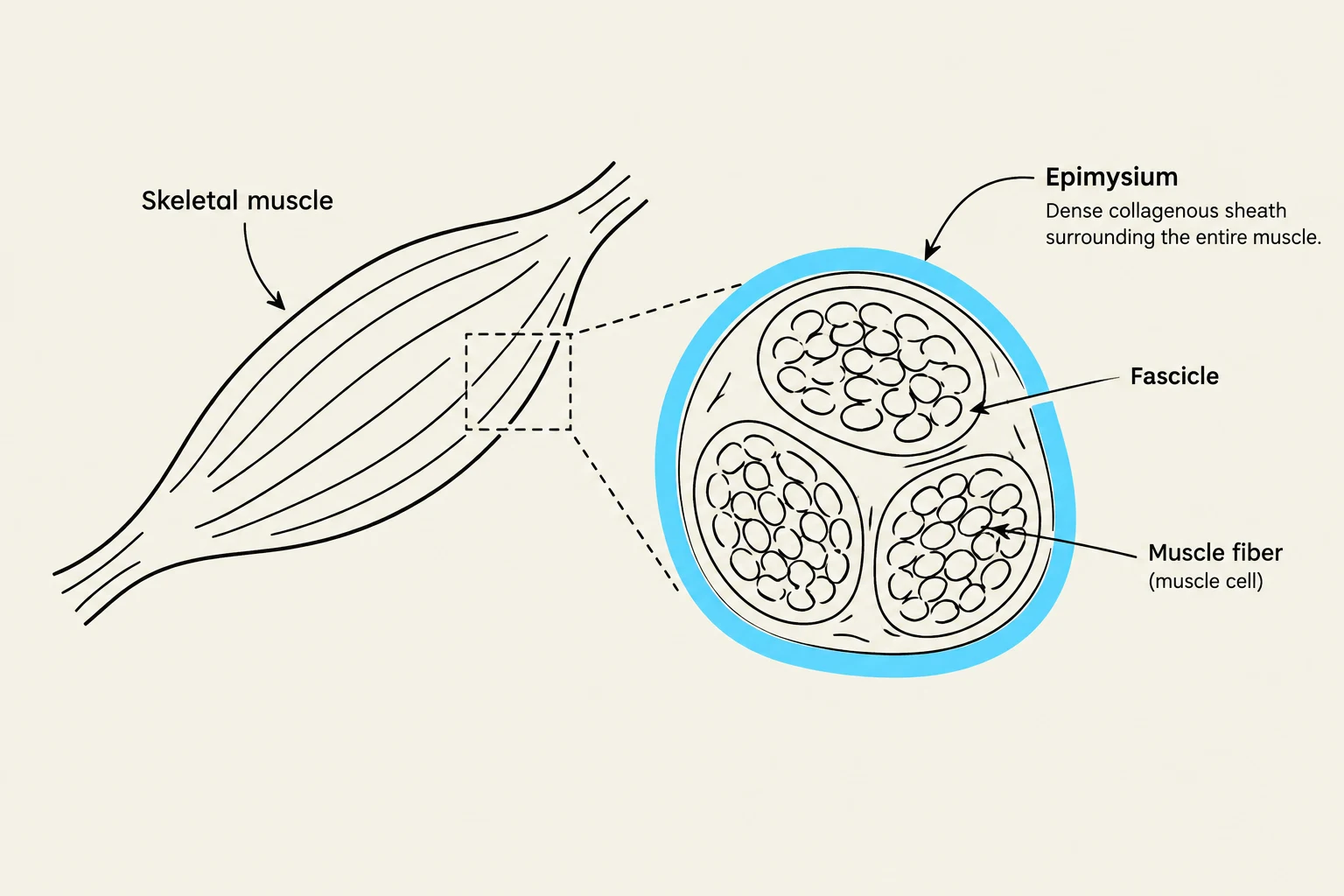
Exam Tip: If a question asks which layer separates a muscle from an adjacent organ, the answer is the epimysium.

Keep reading for free.
Create an account to finish this reading and make it part of your study routine.
Continue reading freeAlready have an account? Log in
Updated