An overview of the cholinergic system, detailing acetylcholine synthesis, release, inactivation, receptor subtypes (nicotinic and muscarinic), and its organization and function in the central and peripheral nervous systems. Includes clinical implications and pharmacology.
The cholinergic system is a fundamental component of both the central and peripheral nervous systems, defined by the use of the neurotransmitter acetylcholine (ACh). This system is responsible for a vast array of physiological and psychological processes, ranging from the basic mechanics of muscle contraction to complex cognitive functions like memory encoding and sustained attention. Understanding how ACh is synthesized, how it interacts with specific receptor subtypes, and how its signaling is regulated is essential for understanding both normal neurobiology and various clinical disorders.
Acetylcholine Synthesis, Release, and Inactivation
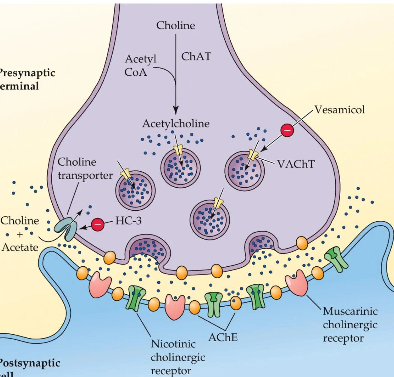
The life cycle of acetylcholine begins with its synthesis within the presynaptic neuron. ACh is synthesized in a single step from two precursors: choline and acetyl coenzyme A (acetyl CoA). This reaction is catalyzed by the enzyme choline acetyltransferase (ChAT), which is found exclusively in cholinergic neurons. The rate of ACh synthesis is not fixed; it depends on the availability of these precursors and the rate of cell firing.
FIGURE 7.1 Synthesis of acetylcholine (ACh) by choline acetyltransferase.
Once synthesized, ACh is loaded into synaptic vesicles by vesicular ACh transporters (VAChT). This process can be disrupted by drugs like vesamicol, which blocks VAChT and reduces the amount of ACh available for release. Following release into the synapse, the signal must be terminated to prevent overstimulation. This is achieved through the rapid breakdown of ACh by the enzyme acetylcholinesterase (AChE).
Diagram illustrating the cholinergic synapse, showing the synthesis, release, and reuptake of acetylcholine, as well as its interaction with nicotinic and muscarinic receptors on the postsynaptic membrane.
FIGURE 7.2 A cholinergic synapse.
AChE breaks ACh down into choline and acetic acid. To ensure the system can continue to function, the resulting choline is recycled back into the neuron via choline transporters. If these transporters are blocked—for example, by the drug hemicholinium-3 (HC-3)—ACh production declines significantly.
Acetylcholinesterase (AChE) Isoforms
AChE is not a uniform enzyme; it exists in different molecular forms depending on its location in the nervous system. These forms are categorized by their subunit structure and how they are anchored to cells:
Soluble G4: A free-floating form consisting of four subunits (a tetramer) within the cell.
Membrane-tethered G4: A four-subunit form anchored to the cell membrane, positioned to allow for rapid breakdown of ACh in the synaptic cleft.
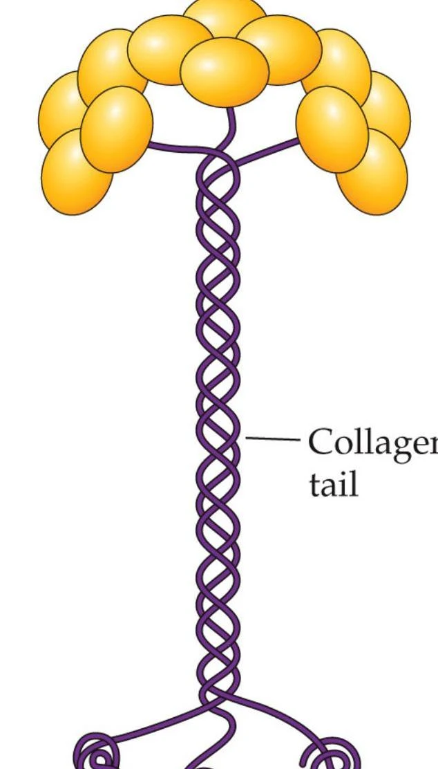
A12 Form: Found specifically at neuromuscular junctions, this form consists of 12 subunits. Its high efficiency allows for the extremely rapid breakdown of ACh, which is critical for allowing muscles to relax quickly before the next contraction.
A diagram showing four yellow spheres connected by a thin black line.
FIGURE 7.4 Soluble G4 form of AChE.
FIGURE 7.4 Membrane-bound G4 form of AChE.
Diagram illustrating the structure of a collagen tail.
FIGURE 7.4 A12 neuromuscular junction form of AChE.
Organization of the Cholinergic System
Cholinergic neurons are distributed throughout both the peripheral and central nervous systems, serving distinct roles in each.
The Autonomic Nervous System (PNS)
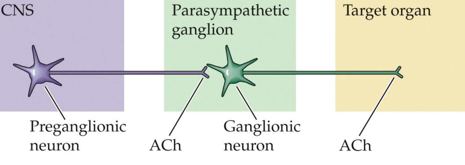
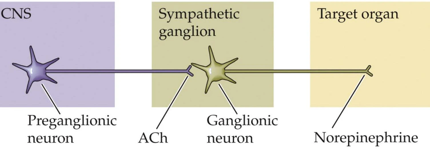
In the peripheral nervous system, cholinergic neurons are essential components of the autonomic pathways. Specifically, the preganglionic neurons of both the sympathetic and parasympathetic branches are cholinergic. Additionally, the ganglionic neurons of the parasympathetic system utilize ACh.
Diagram illustrating the pathway of a parasympathetic neuron from the CNS to a target organ, showing the preganglionic and ganglionic neurons and the neurotransmitter acetylcholine (ACh).
FIGURE 7.7 Parasympathetic branch.
Diagram illustrating the pathway of a sympathetic neuron from the CNS to a target organ, involving a preganglionic neuron, sympathetic ganglion, and the release of norepinephrine.
FIGURE 7.7 Sympathetic branch.
The Central Nervous System (CNS)
In the brain, cholinergic neurons are clustered in specific nuclei that project to various regions.
Basal Forebrain Cholinergic System (BFCS): This system provides critical innervation to the cerebral cortex and the hippocampus. Projections from the medial septum and diagonal band to the hippocampus are involved in the encoding of declarative memories, while projections from the nucleus basalis to the prefrontal cortex are vital for maintaining sustained attention.
LDTg and PPTg Nuclei: These nuclei project to the basal ganglia and thalamus. They play diverse roles, including the initiation of REM sleep and the stimulation of midbrain dopamine neurons, which is a key component of drug reward and reinforcement.
Striatum: Interneurons in the striatum help regulate movement through a delicate balance between ACh and dopamine.
Clinical Note: Parkinson's Disease
In Parkinson's disease, a loss of dopamine (DA) creates a transmitter imbalance in the striatum. Because of this imbalance, anticholinergic drugs (which reduce ACh activity) may be used as part of a treatment strategy.
Receptor Subtypes: Nicotinic and Muscarinic
ACh exerts its effects through two primary classes of receptors: ionotropic (nicotinic) and metabotropic (muscarinic).
Nicotinic Acetylcholine Receptors (nAChRs)
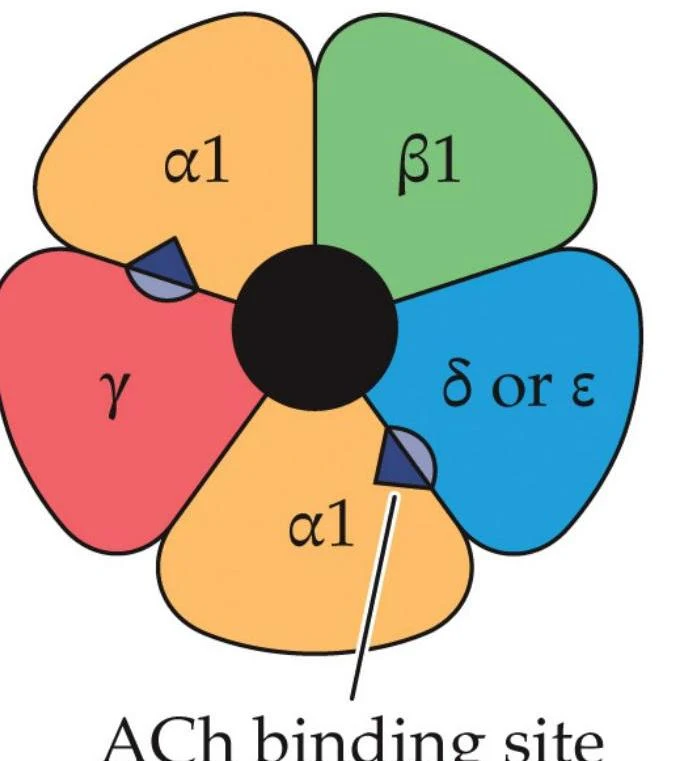
Nicotinic receptors are ionotropic, meaning they function as ligand-gated ion channels that mediate fast excitatory responses. When ACh binds, the channel opens, allowing Na+ and Ca2+ to flow into the cell, causing depolarization. These receptors are pentameric, composed of five subunits (such as α,β,γ,δ, and ε).
Diagram illustrating the subunits of a nicotinic acetylcholine receptor, including alpha1, beta1, gamma, and delta or epsilon subunits, with a labeled acetylcholine binding site.
FIGURE 7.10 Muscle nicotinic receptor.
Nicotinic receptors exist in three functional states: closed, open, or desensitized. In the desensitized state, the channel remains closed even if the agonist is bound. Furthermore, continuous exposure to an agonist can lead to a "depolarization block," where the membrane remains so depolarized that it loses its resting potential and cannot be excited again until the agonist is removed.
Muscarinic Acetylcholine Receptors (mAChRs)
Muscarinic receptors are metabotropic, meaning they operate through second-messenger systems rather than direct ion channels. There are five subtypes categorized by their effect on cellular signaling:
Excitatory (M1,M3,M5): These subtypes activate the phosphoinositide second-messenger system.
Inhibitory (M2,M4): These subtypes inhibit the synthesis of cAMP and stimulate the opening of K+ channels.
In the brain, muscarinic receptors in the neocortex and hippocampus are essential for cognitive functions. Outside the brain, they are found in cardiac and smooth muscle, where they are activated by the parasympathetic nervous system.
Clinical Implications and Pharmacology
Dysfunction in the cholinergic system is linked to several significant medical conditions:
Myasthenia Gravis: An autoimmune disorder where the body produces antibodies that block muscle nicotinic receptors. This leads to severe muscle weakness and fatigue. It is often treated with drugs like neostigmine or pyridostigmine, which are AChE inhibitors that increase the availability of ACh at the neuromuscular junction.
Dementia: Age-related dementia is often treated with reversible AChE inhibitors, such as donepezil (Aricept), which increase ACh levels in the brain to improve cognitive function.
Pharmacological agents can also target these systems for other purposes:
AChE Inhibitors: While reversible inhibitors like donepezil are used for dementia, irreversible inhibitors like organophosphorus compounds (used as insecticides or nerve gases) can be fatal by causing massive ACh accumulation.
nAChR Antagonists: Bubocurarine (the active ingredient in curare) acts as an antagonist at muscle nicotinic receptors, causing paralysis.
Exam Tip: Receptor States
Remember that "desensitization" is a functional state where the receptor is bound by an agonist but the channel remains closed. This is distinct from a "depolarization block," which refers to the inability of the membrane to repolarize due to persistent activation.
Keep reading for free.
Create an account to finish this reading and make it part of your study routine.