Explores the structural and functional organization of the nervous system, including the central and peripheral nervous systems, and the cellular components: neurons and glial cells.
The nervous system is a complex, highly organized network responsible for coordinating almost every function in the human body. It operates by detecting environmental changes, processing that information, and initiating appropriate responses. To understand how this system works, we must look at it through two different lenses: its structural organization (where the parts are located) and its functional organization (how the information flows).
Structural Organization: CNS and PNS
Structurally, the nervous system is divided into two primary subdivisions:
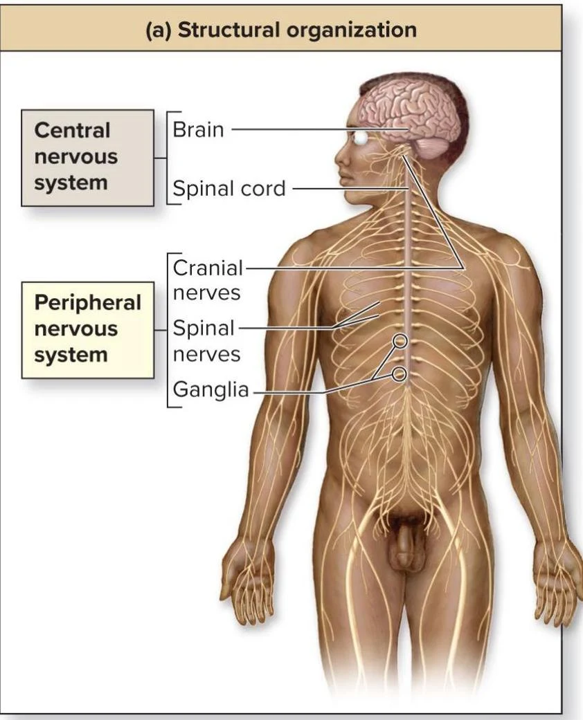
Central Nervous System (CNS): This serves as the control center and consists of the brain and the spinal cord.
Peripheral Nervous System (PNS): This consists of the nerves that extend from the CNS to the rest of the body, including the cranial nerves, spinal nerves, and ganglia.
This illustration shows the structural organization of the human nervous system, differentiating between the central and peripheral nervous systems, and labeling key components like the brain, spinal cord, cranial nerves, spinal nerves, and ganglia.
Figure 14.1a
Functional Organization: The Flow of Information
Functionally, the nervous system is organized based on the direction in which information travels. This creates a continuous cycle of input, processing, and output.
The Sensory (Afferent) Nervous System
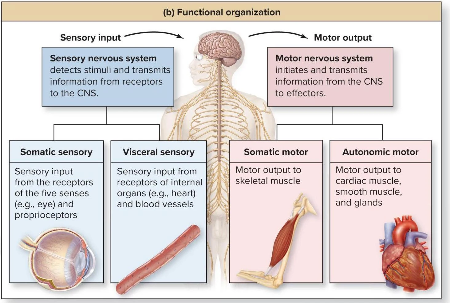
The sensory nervous system is responsible for "input." It collects information from receptors located in the PNS and transmits it to the CNS. This system is further divided into two categories:
Somatic sensory: This involves the conscious perception of general senses, such as touch, pain, pressure, vibration, and proprioception (the sense of body position).
Visceral sensory: These are impulses that are generally unconscious, originating from the internal organs (viscera) and blood vessels.
The Motor (Efferent) Nervous System
The motor nervous system is responsible for "output." It carries impulses from the CNS through the PNS to effectors, such as muscles or glands. It is divided into:
Somatic motor (Somatic Nervous System; SNS): This governs voluntary control, specifically by conducting impulses that cause the contraction of skeletal muscles.
Autonomic motor (Autonomic Nervous System; ANS): Also known as the visceral motor system, this governs involuntary functions by regulating smooth muscle, cardiac muscle, and glands.
This diagram illustrates the functional organization of the nervous system, differentiating between sensory input and motor output pathways, and categorizing them into somatic and visceral sensory, and somatic and autonomic motor functions.
Figure 14.1b
Cytology: The Cells of Nervous Tissue
At the cellular level, nervous tissue is composed of two distinct types of cells: neurons and glial cells.
Neurons: The Communicators
Neurons are the basic structural and functional units of the nervous system. They are electrically excitable cells that initiate, transmit, and receive nerve impulses (rapid movements of an electrical charge).
Neurons possess several unique characteristics:
Conductive and Excitable: They propagate electrical signals in response to stimuli.
Secretory: They release neurotransmitters to communicate with other cells.
Metabolic and Long-lived: They require abundant glucose and oxygen and can remain functional from fetal development into very old age.
Amitotic: Most neurons lose the ability to undergo cell division once they are formed.
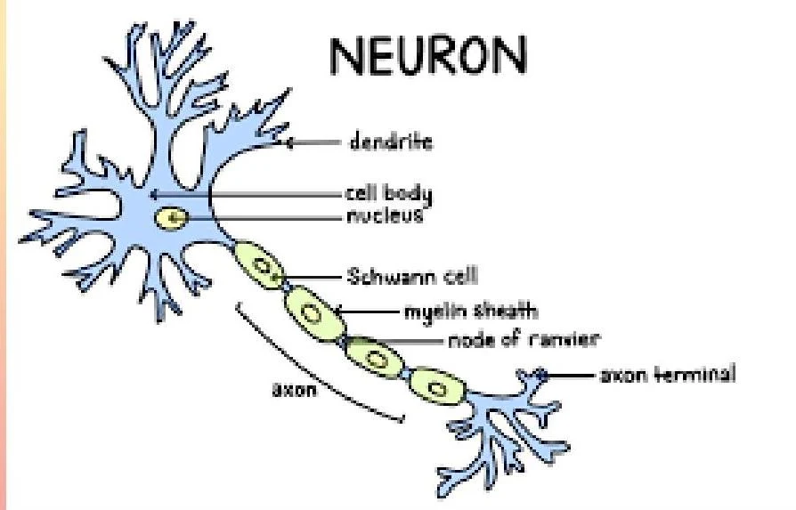
A typical neuron consists of a cell body (soma or perikaryon), dendrites (short processes that receive signals), and an axon (a long process that transmits impulses away from the cell body).
Diagram of a neuron, illustrating its key components such as dendrites, cell body, nucleus, axon, Schwann cells, myelin sheath, nodes of Ranvier, and axon terminals.
Figure 14.2
Neurons can be classified structurally (unipolar, bipolar, or multipolar) and functionally (sensory, motor, or interneurons).
Glial Cells: The Support System
Glial cells (or neuroglia) are non-excitable cells that provide support, nourishment, and protection for neurons. While they do not typically transmit nerve impulses, they are more numerous than neurons and are capable of mitosis.
Glial Cells of the CNS
There are four main types of glial cells in the central nervous system:
Astrocytes: The most abundant type; they help form the blood-brain barrier (BBB), regulate tissue fluid, and assist in neuronal development.
Ependymal cells: Ciliated cells that line the ventricles of the brain and the central canal of the spinal cord, helping to produce cerebrospinal fluid (CSF).
Microglial cells: Act as the immune defense by removing debris and dead or damaged tissue.
Oligodendrocytes: These cells wrap their processes around CNS axons to form the myelin sheath, an insulating layer that increases the speed of impulse conduction.
Glial Cells of the PNS
In the peripheral nervous system, two types of glial cells predominate:
Satellite cells: These regulate the fluid environment around neuron cell bodies in the PNS ganglia.
Neurolemmocytes (Schwann cells): These are responsible for forming the myelin sheath around axons in the PNS.
Exam Tip: Remember the distinction in myelination! Oligodendrocytes myelinate many axons in the CNS, whereas a single neurolemmocyte wraps around only one segment of a single axon in the PNS.
Myelination and Impulse Conduction
Myelination is the process of wrapping an axon in a lipid-rich sheath to provide electrical insulation. This insulation significantly changes how nerve impulses travel.
Saltatory Conduction: In myelinated axons, the impulse appears to "jump" from one node (an exposed region of the membrane) to the next. This is a very rapid form of conduction.
Continuous Conduction: In unmyelinated axons, the impulse must travel the entire length of the axon membrane, which is a much slower process.
Clinical Note: Because neurolemmocytes in the PNS form a protective sheath called the neurilemma, damaged PNS axons have the potential to regenerate if the cell body remains intact. This is much more difficult in the CNS.
Keep reading for free.
Create an account to finish this reading and make it part of your study routine.