


The physical assessment of the ear is a systematic process that combines careful inspection and palpation of external structures with a detailed otoscopic examination of the ear canal and tympanic membrane. Because the ear is integral to both hearing and equilibrium, a thorough examination is essential for identifying infections, structural abnormalities, and neurological implications.
Subjective Data and Clinical History
Before beginning the physical exam, clinicians must gather a comprehensive history to guide their objective findings. Key areas of inquiry include:
- Earaches and Infections: Assess the nature of the pain (e.g., sharp, dull, or throbbing) and inquire about recent upper respiratory infections (URIs), which can impact middle ear pressure.
- Discharge (Otorrhea): Determine if any fluid is exiting the canal, which may indicate infection or a ruptured eardrum.
- Tinnitus: Ask about the perception of ringing, buzzing, or other noises.
- Vertigo: Inquire about spinning or whirling sensations. This often indicates inflammation of the labyrinth, the inner ear structure that acts as a biological "plumb line" for equilibrium.
- Hearing Loss and Recruitment: Specifically ask about recruitment, a condition where low-intensity sounds become distorted and painfully loud when repeated.
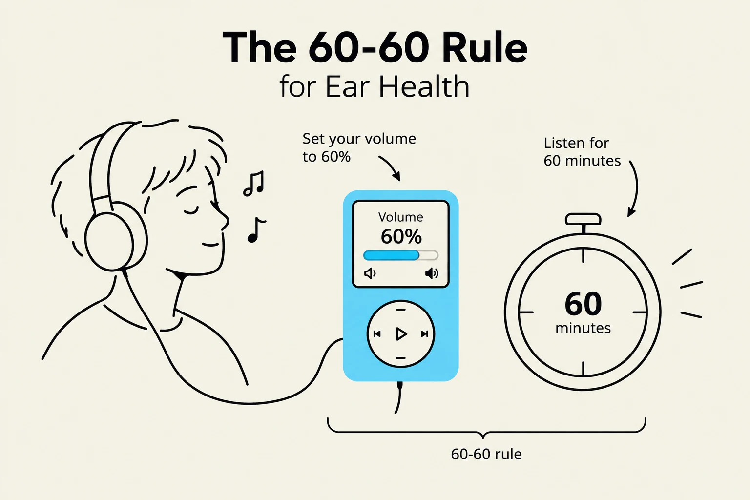
- Noise Exposure: Identify risks from loud environments or the use of earbuds, which deliver excessive sound pressure directly into the canal.
External Ear: Inspection and Palpation
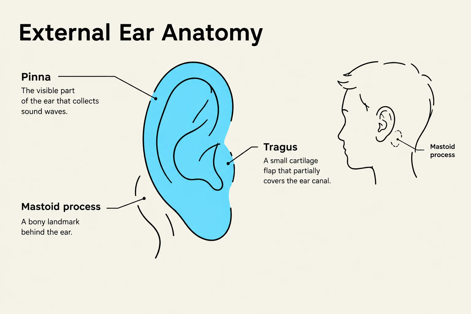
The objective exam begins with the external ear, focusing on the pinna (auricle) and the surrounding skin.
Size, Shape, and Skin Condition
The ears should be bilaterally equal in size and shape. While variations can be familial, certain findings are clinically significant:
- Microtia: An underdeveloped pinna or auricle.
- Macrotia: An abnormally large ear, defined by a height and a width .
- Skin Lesions: Clinicians should look for tophi (hard, whitish-yellow uric acid nodules from gout), sebaceous cysts, keloids, and carcinoma (ulcerated, bleeding nodules with an indurated base).
- Discoloration: Red-blue discoloration may indicate frostbite.
- Crusting/Scaling: This may suggest otitis externa (Swimmer's Ear), eczema, or seborrhea.
Keep reading for free.
Create an account to finish this reading and make it part of your study routine.
Continue reading freeAlready have an account? Log in
Updated