This guide details the nursing care for patients with tracheostomies, covering insertion, post-procedure monitoring, airway patency, emergency management of decannulation, and long-term care including speech, swallowing, and home ventilation.
A tracheostomy is a surgically created opening (stoma) in the anterior surface of the neck designed to establish a patent airway, bypass upper airway obstructions, or facilitate the removal of secretions. While often used in emergency situations, it is primarily indicated for patients requiring long-term mechanical ventilation (typically >2 weeks). For the nurse, managing a tracheostomy patient requires a high level of vigilance, ranging from immediate post-procedure monitoring to the complex physiological management of long-term home ventilation.
Tracheostomy Insertion and Post-Procedure Care
Tracheostomy procedures may occur in the operating room or at the bedside. When performed at the bedside, the nurse must ensure that emergency resuscitation equipment, a working bag-valve-mask (BVM), and suctioning equipment are immediately available.
During the procedure, the nurse monitors hemodynamic status, oxygen saturation (SpO2), and the patient's response to sedation. Immediately following insertion, the nurse must confirm tube placement by auscultating for bilateral breath sounds, observing symmetric chest wall movement, and utilizing EtCO2 capnography.
Post-Procedure Monitoring Priorities
Once the procedure is complete, the following nursing interventions are critical:
Vital Signs and Oxygenation: Continuously monitor SpO2 and respiratory rate.
Radiology: Obtain a chest X-ray to confirm the exact location of the tube.
Site Assessment: Monitor for bleeding at the insertion site and assess for signs of infection.
Ventilator Settings: Document the mode, fraction of inspired oxygen (FiO2), and Positive End-Expiratory Pressure (PEEP).
A post-procedure checklist illustrates three steps: confirming placement via auscultation and EtCO2, obtaining a chest X-ray (CXR) to check tube position and lungs, and monitoring SpO2 for oxygenation.
Maintaining Airway Patency and Safety
The two most critical components of tracheostomy safety are the cuff and the ties.
Tracheostomy Cuff Pressure
Most tracheostomy tubes used in mechanically ventilated patients are cuffed. The cuff is an inflatable sleeve that seals the trachea to prevent the escape of ventilating gases and protects against aspiration. However, improper pressure management can lead to complications:
Too much pressure: May damage the tracheal mucosa.
Too little pressure: May lead to an audible cuff leak or inadequate ventilation.
Nursing Alert: Use the least amount of air needed to maintain an airway seal. Measure cuff inflation pressure with a manometer at least every 8 hours. Pressure should generally not exceed 20 to 30 cm H2O (15–22 mmHg).
Stoma Care and Trach Tie Replacement
To prevent infection and skin breakdown, the nurse must perform sterile dressing changes every 12–24 hours. Additionally, the tracheostomy ties (tapes) must be managed carefully to ensure the tube remains secure without causing skin irritation.
When replacing trach ties, always use two people: one person stabilizes the tracheostomy flange to prevent dislodgement, while the second person changes the tapes. It is best practice to tie the tapes securely with enough room to fit two fingers between the tape and the patient's skin.
Suctioning Protocols
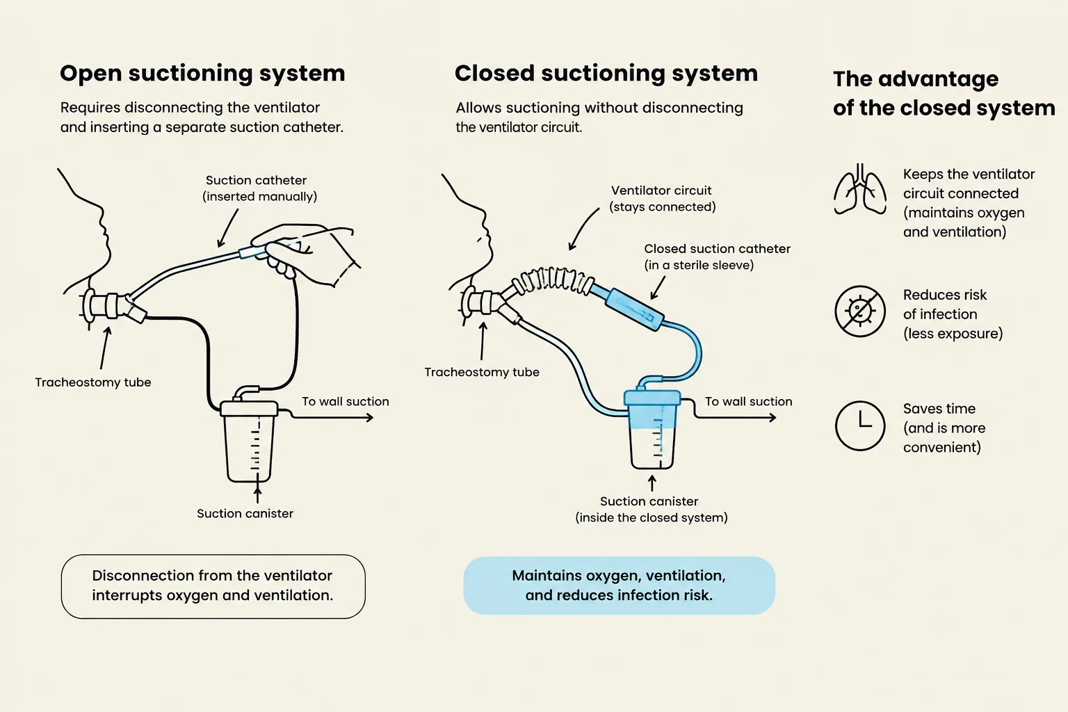
Suctioning is necessary to maintain tube patency, but it should not be performed routinely. Instead, it should be performed based on clinical need.
Indications for suctioning include:
Visible secretions in the tracheostomy tube.
Sudden decrease in SpO2.
Increased respiratory rate or frequent coughing.
Auscultation of adventitious breath sounds over the trachea.
Comparison of open and closed suctioning systems for tracheostomy tubes, highlighting the advantages of the closed system in maintaining ventilation and reducing infection risk.
Emergency Management: Accidental Decannulation
Accidental decannulation is a life-threatening emergency where the tracheostomy tube is dislodged.
Immediate Nursing Actions:
Call for help immediately.
Assess the patient: Check level of consciousness (LOC), ability to breathe, and signs of respiratory distress.
Positioning: Place the patient in a semi-Fowler’s position.
Ventilation: Ventilate the patient with a BVM over the nose and mouth. If the patient has undergone a laryngectomy, you must ventilate through the stoma.
Stoma Protection: Cover the stoma with a sterile dressing.
Decannulation and Long-Term Management
The Decannulation Procedure
Decannulation is the intentional removal of the tracheostomy tube once the patient no longer requires it. A patient is a candidate for decannulation if they are hemodynamically stable, have a stable respiratory drive, can exchange air adequately, and can independently expectorate secretions.
The procedure involves:
Suctioning the patient and clearing oral secretions.
Deflating the cuff and removing the tube in one smooth motion.
Applying a sterile occlusive dressing to the stoma.
Long-Term Care and Communication
For patients with long-term tracheostomies, the focus shifts to rehabilitation and quality of life.
Speech and Swallowing:
Patients with long-term tracheostomies may face swallowing difficulties and a risk for aspiration. A Speech-Language Therapist (SLT) should assess swallowing ability. If a patient can swallow without aspiration, a cuffless tube may be used. To facilitate communication, a Passy-Muir valve may be used. This device requires the patient to exhale through the nose and mouth instead of through the tracheostomy, allowing for speech.
Home Mechanical Ventilation:
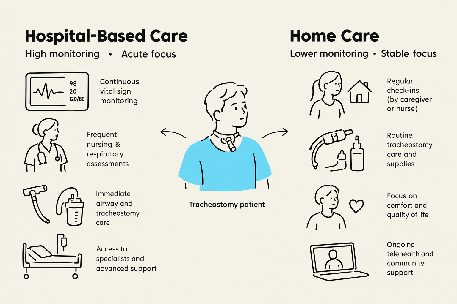
Some patients may transition to home mechanical ventilation. This can be achieved using negative pressure ventilators (which do not require an artificial airway) or positive pressure ventilators. While home care allows for greater mobility and family integration, nurses must educate caregivers on equipment troubleshooting, recognizing low supplies, and managing caregiver fatigue.
This illustration compares hospital-based care with home care for a tracheostomy patient, highlighting key differences in monitoring, focus, and available support.
Exam Tip: When deciding whether to suction a patient, remember that "routine" suctioning is not recommended. Always assess for clinical indicators like increased peak airway pressure or decreased SpO2 first.
Keep reading for free.
Create an account to finish this reading and make it part of your study routine.