Nursing Management of Pregnancy-Related Complications
This article explores the primary categories of pregnancy-related risks, focusing on metabolic, cardiac, infectious, and obstetric challenges. It details nursing management strategies for conditions such as diabetes mellitus, cardiac disease, infections, and labor alterations.
Pregnancy is a period of significant physiological adaptation, but it can also introduce serious health risks that threaten the well-being of both the mother and the fetus. Effective nursing management requires a proactive approach: identifying high-risk conditions through meticulous assessment, implementing targeted interventions, and providing continuous surveillance to mitigate complications. This article explores the primary categories of pregnancy-related risks, focusing on metabolic, cardiac, infectious, and obstetric challenges.
Diabetes Mellitus in Pregnancy
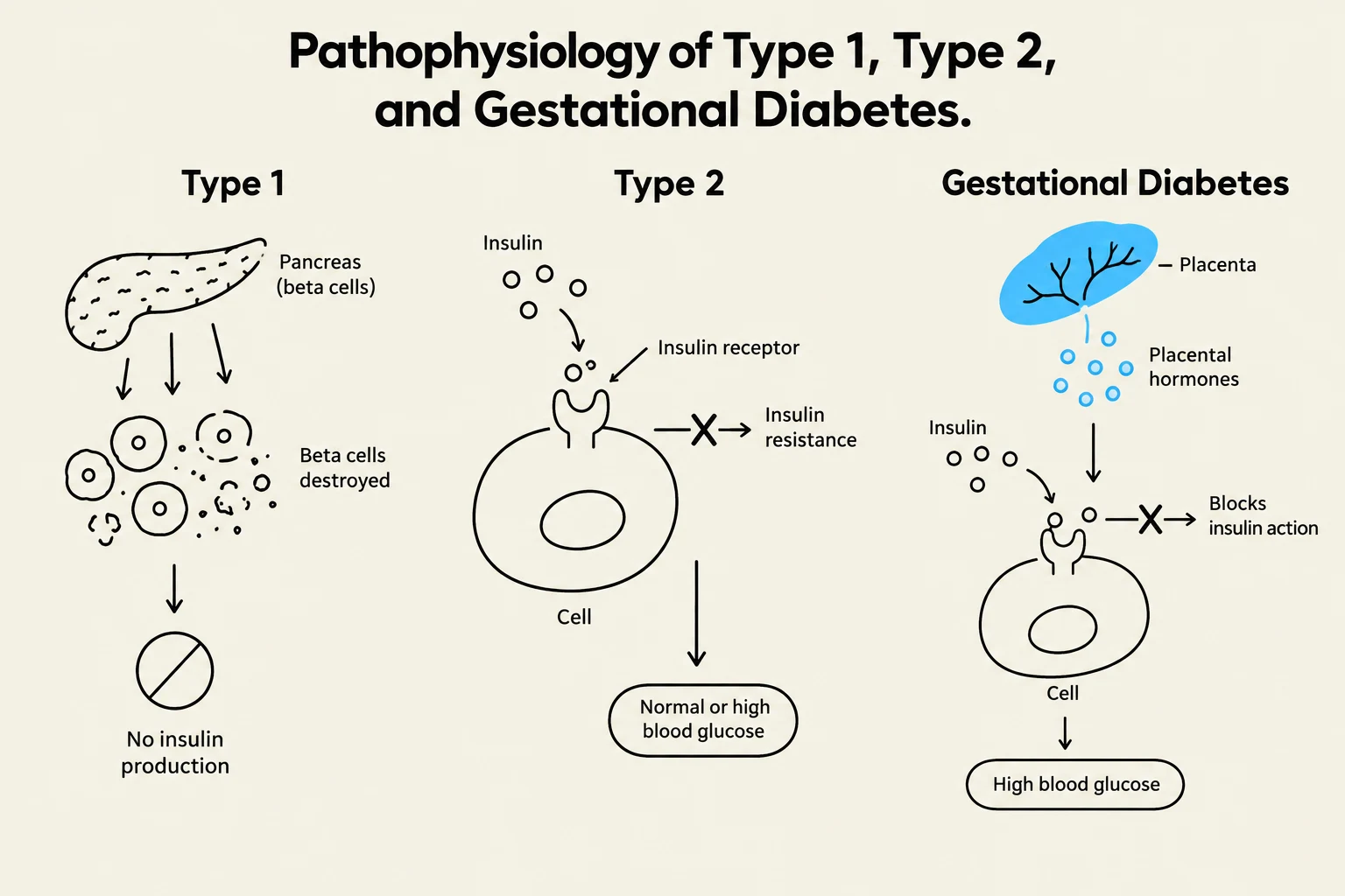
Diagram illustrating the pathophysiology of Type 1, Type 2, and Gestational Diabetes, showing the mechanisms of insulin production and action in each condition.
Diabetes mellitus is a major concern in prenatal care, categorized by its onset and underlying pathophysiology.
Type 1 and Type 2 Diabetes
Pre-gestational diabetes refers to women who have diabetes prior to conception.
Type 1 Diabetes: Characterized by an absolute insulin deficiency caused by the destruction of pancreatic beta cells.
Type 2 Diabetes: Involves insulin resistance or a progressive loss of beta cell secretion.
Because most major structural defects in the renal, cardiac, skeletal, and CNS systems occur by the eighth week of gestation, preconception counseling is vital for women with these conditions.
Gestational Diabetes Mellitus (GDM)
GDM is a carbohydrate intolerance that results in hyperglycemia, typically manifesting in the second or third trimester due to placental hormones. While some women can compensate for hormonal changes by producing more insulin, those with GDM cannot, leading to elevated blood glucose levels.
Screening and Diagnosis
Nurses must screen patients at their initial visit for risk factors such as obesity, hypertension, glycosuria, and a history of delivering an infant >4,000 g (approx. 9 lbs).
1-Hour Glucose Tolerance Test (Screening): A 50g oral glucose load is administered (fasting is not required). If the result is >140 mg/dL, a follow-up 3-hour test is necessary.
3-Hour Glucose Tolerance Test (Diagnostic): This requires the patient to be fasting and involves a 100g oral glucose load. A diagnosis of GDM is confirmed if one or more of the following thresholds are exceeded:
Fasting: ≥95 mg/dL
1-hour: ≥180 mg/dL
2-hour: ≥155 mg/dL
3-hour: ≥140 mg/dL
Nursing Management of GDM
The foundation of treatment is nutritional management using a restricted carbohydrate diet. Patients must be educated not to attempt weight loss or enter ketosis during this period. If diet and exercise are insufficient, insulin is the preferred pharmacological treatment. Oral hypoglycemics like glyburide are also considered safe because they do not cross the placenta.
Nursing Surveillance Tip:
Nurses should monitor maternal HbA1c every 4–6 weeks, check urine for protein and ketones, and perform fetal surveillance via ultrasound, nonstress testing (NST), and biophysical profiles.
Complications
Maternal Complications
Neonatal Complications
Hypertension
Macrosomia (large birth weight)
Preeclampsia
Hypoglycemia
Increased risk of Cesarean birth
Birth trauma (e.g., shoulder dystocia)
Future risk of Type 2 DM
Respiratory distress syndrome
During labor, nurses should aim to keep maternal glucose levels stable around 110 mg/dL and monitor the neonate closely for hypoglycemia immediately after delivery.
Cardiac Disease in Pregnancy
Pregnancy causes significant hemodynamic changes that can overstress the cardiovascular system.
Congenital Heart Disease: Structural defects present from birth.
Acquired Heart Disease: Developed during the patient's lifetime.
High-risk populations include non-Hispanic Black women, those over age 40, and those with obesity or hypertension. Nurses must assess for signs of cardiac decompensation, such as dyspnea, cyanosis, or palpitations.
Management Strategies:
Hemodynamic Stability: Prevent sudden drops in maternal blood pressure or volume, which can shunt blood away from the uterus and reduce placental perfusion.
Energy Conservation: Encourage rest in the side-lying position and prioritize rest periods.
Nutrition: High-fiber diet to prevent straining/constipation and adequate hydration.
Medications: Use of Lasix, Digitalis, or Heparin may be prescribed; however, Warfarin is contraindicated.
Infections
Infections can have devastating effects on both the mother and the developing fetus.
HIV: Risk of vertical transmission via placenta or breastmilk. With appropriate Antiretroviral Therapy (ART), transmission risk drops from 35% to <1%. Breastfeeding is not recommended.
Group B Streptococcus (GBS): A common bacterium in the GI/GU tract. To prevent neonatal sepsis or meningitis, patients are tested at 36–37 weeks; if positive, IV Penicillin G is administered at least 4 hours before birth.
TORCH Infections: Includes Toxoplasmosis (from raw meat/cat feces) and Cytomegalovirus (CMV), which is a major cause of intrauterine growth restriction (IUGR).
Syphilis: Can cause spontaneous abortion, stillbirth, or neonatal bone damage.
Hepatitis B: If the mother is positive, the newborn must receive Hepatitis B Immune Globulin (HBIG) and the Hep B vaccine within 12 hours of birth.
Herpes Simplex Virus (HSV): If active lesions are present within 6 weeks of term, a Cesarean section is recommended to avoid contact with the virus during delivery.
Alterations in Labor and Delivery
Dystocia and Shoulder Dystocia
Dystocia is the abnormal, prolonged progression of labor, often leading to Cesarean sections. It can be caused by problems with the "powers" (contractions), the "passenger" (fetus), the "passageway" (pelvis), or the mother's "psyche" (anxiety/pain).
Shoulder Dystocia is an obstetric emergency where the fetal shoulder is obstructed by the maternal pelvis after the head has been delivered.
Sign: The "Turtle Sign" (fetal head retracts toward the perineum).
Nursing Intervention: Call for help immediately and initiate the McRoberts maneuver (flexing and abducting the mother's thighs) followed by suprapubic pressure. Do not apply traction to the head.
Preterm Labor
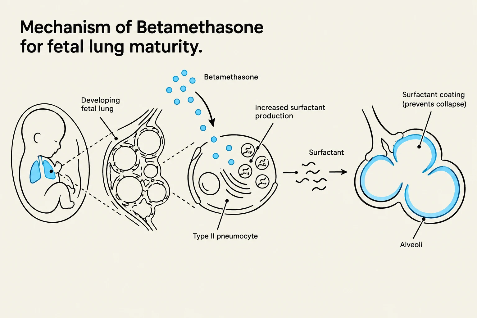
This diagram illustrates the mechanism by which Betamethasone promotes fetal lung maturity by increasing surfactant production in Type II pneumocytes, which prevents alveolar collapse.
Preterm labor is defined as regular uterine contractions with cervical effacement and dilation occurring between 20 and 37 weeks of gestation.
Management includes:
Tocolytics: Medications to slow or stop contractions (e.g., Magnesium Sulfate, Indomethacin, Nifedipine).
Fetal Lung Maturity: Administration of Betamethasone (2 doses, 24 hours apart) between 24–34 weeks to stimulate surfactant production.
Induction, Augmentation, and Assisted Birth
Induction/Augmentation: Stimulation of contractions via cervical ripening (prostaglandins) or Pitocin (synthetic oxytocin).
Instrumental Delivery: Use of a vacuum (suction) or forceps (tongs) to assist the fetal head during the second stage of labor.
Cesarean Birth: Indicated for reasons such as placenta previa, active herpes, or fetal distress. Postoperative nursing care focuses on monitoring vital signs, lochia, and surgical sites.
Keep reading for free.
Create an account to finish this reading and make it part of your study routine.