Nursing Management of Pediatric Respiratory Infections: Pertussis and Tuberculosis
This article details the nursing considerations for managing Pertussis (Whooping Cough) and Tuberculosis (TB) in pediatric patients, covering their distinct pathophysiological pathways, clinical stages, diagnostic evaluations, and essential nursing interventions for patient safety.
Respiratory infections in the pediatric population require vigilant nursing assessment and specialized management due to the unique physiological characteristics of children. Compared to adults, children have smaller airway diameters, which increases the risk of obstruction, and highly vascular mucous membranes that are susceptible to edema and spasm. This article explores the nursing considerations for two significant infectious diseases: Pertussis (Whooping Cough) and Tuberculosis (TB), focusing on their distinct pathophysiological pathways and the clinical management required to ensure patient safety.
Pertussis (Whooping Cough)
Pertussis is an acute respiratory tract infection caused by the bacterium Bordetella pertussis. It is highly contagious and is transmitted through direct contact or droplet spread. While it can affect all ages, it is particularly life-threatening for infants under four years old and those who are unimmunized.
Pathophysiology of B. pertussis
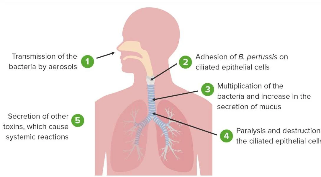
The infection begins when B. pertussis enters the respiratory tract via the nose or mouth and attaches to the ciliated epithelial cells in the upper airways. Once attached, the bacteria release toxins that lead to:
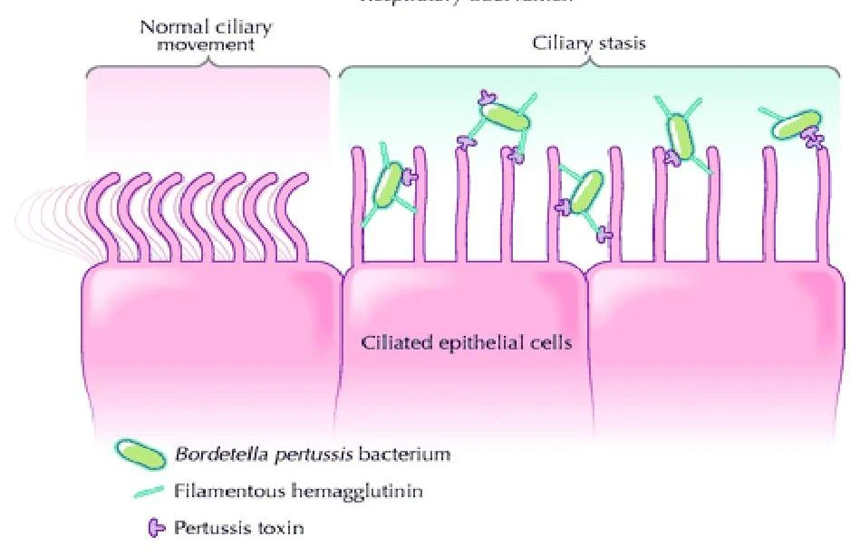
Ciliary dysfunction (stasis).
Excessive mucous production.
Immune suppression, specifically affecting the function of neutrophils and macrophages through high levels of cyclic AMP.
Diagram illustrating the stages of Bordetella pertussis infection in the respiratory system, including transmission, adhesion to epithelial cells, bacterial multiplication, paralysis of ciliated cells, and toxin secretion.
Illustration showing Bordetella pertussis bacteria adhering to ciliated epithelial cells, leading to ciliary stasis.
Clinical Stages
Pertussis typically progresses through three distinct stages:
Catarrhal Stage (1–2 weeks): The most contagious phase. Symptoms are mild and resemble a common cold, including sneezing, nasal congestion, and a low-grade fever. In infants, this stage may present as apnea rather than a cough.
Paroxysmal Stage (2–6+ weeks): Characterized by violent coughing fits (paroxysms) often followed by a high-pitched "whoop" sound. Patients may experience cyanosis, respiratory failure, and post-tussive vomiting.
Convalescent Stage (1–2 weeks): Symptoms gradually decrease in frequency and severity, though a persistent cough may remain for several weeks.
Nursing Management of Pertussis
Nursing care focuses on airway protection, respiratory support, and hydration.
Assessment: Conduct frequent, comprehensive cardiorespiratory and neurological assessments. Monitor oxygen saturation and airway patency closely.
Airway & Respiratory Support: Administer humidified supplemental oxygen as ordered. Suction the airway as needed, but ensure the patient is kept in a side-lying position to prevent aspiration, especially if post-tussive vomiting occurs.
Nutrition & Hydration: Ensure adequate hydration via oral or IV fluids. For infants, provide small, frequent feeds or continuous enteral feeds to manage the risk of vomiting.
Infection Control: Implement droplet precautions and facilitate contact tracing.
Psychosocial Support: Provide emotional support to caregivers, who may experience guilt regarding immunization status.
Exam Tip: When assessing an infant with suspected pertussis, remember that they may not present with the classic "whoop." Instead, look for episodes of apnea, which can be a primary clinical manifestation in this age group.
Tuberculosis (TB)
Tuberculosis is an infectious disease caused by Mycobacterium tuberculosis (or the bovine strain). It is transmitted via airborne droplets that can remain suspended in the air for several hours.
This diagram illustrates the factors influencing exposure risk, including proximity, time spent in a shared space, and ventilation. Increased proximity, longer duration, and poor ventilation are associated with higher transmission risk.
Diagram illustrating the transmission of tuberculosis through respiratory droplets containing Mycobacterium tuberculosis.
Pathophysiology and Immune Response
The progression of TB is heavily dependent on the host's immune response:
Inhalation & Phagocytosis:M. tuberculosis enters the lungs via inhaled droplets and travels to the alveoli. Alveolar macrophages and neutrophils attempt to engulf the bacilli via phagocytosis.
Replication & Inhibition: The bacilli replicate inside the macrophages by inhibiting the fusion of the phagosome and lysosome, allowing them to survive and multiply.
Granuloma Formation: The immune system attempts to "wall off" the infection by forming a granuloma (or tubercle).
Progression:
If the infection is contained within the granuloma, it results in Latent TB.
If the infection is not contained, it can spread to lymph nodes or the bloodstream, leading to Active TB disease.
This illustration depicts the pathogenesis of tuberculosis (TB), showing how Mycobacterium tuberculosis (M.tb) enters the lungs, is phagocytosed by alveolar macrophages, and can spread to other organs, causing various complications such as hepatitis, meningitis, and renal parenchyma fibrosis.
Latent vs. Active TB Disease
It is critical to differentiate between these two states for treatment and isolation purposes:
Feature
Latent TB Infection (LTBI)
Active TB Disease
Symptoms
Asymptomatic; no physical findings
Persistent cough, fever, night sweats, weight loss, malaise
Contagiousness
Not infectious
Infectious (via airborne droplets)
Imaging (CXR)
Normal or evidence of healed infection (calcification)
May show lymphadenopathy, pleural effusion, or lung cavitation
Skin Test (TST)
Positive
Positive
Pediatric Considerations: Paucibacillary Disease
Diagnosing TB in children is uniquely challenging. Children under 10 often present with non-specific or mild symptoms and are frequently unable to produce sputum. Furthermore, children often present with paucibacillary disease, meaning they have a low number of bacteria in their system. This makes laboratory confirmation difficult, as fewer than 40% of pediatric cases may be culture-positive.
Diagnostic Evaluation
Tuberculin Skin Test (TST/Mantoux): A type IV hypersensitivity reaction detected 2 to 10 weeks after infection. A positive result requires clinical follow-up and a chest X-ray (CXR). Note that the BCG vaccine can cause a false positive.
IGRA (Quantiferon-TB): An interferon-gamma release assay that confirms exposure and is not affected by the BCG vaccine.
Bacteriologic Exam: Culturing sputum or performing gastric lavage (especially in children who cannot expectorate) to identify acid-fast bacilli (AFB).
Radiography: CXR is used to look for hilar adenopathy or lung opacities.
Nursing Management of Tuberculosis
Management involves a combination of pharmacologic therapy, nutritional support, and strict infection control.
Infection Control: Implement airborne precautions. For patients with smear-positive sputum, isolation must continue until they have provided at least three consecutive smear-negative samples.
Medication Administration:
Latent TB: Typically treated with Isoniazid (INH) for 9 months.
Active TB: Requires a multi-drug regimen (Isoniazid, Rifampin, Ethambutol, and Pyrazinamide).
Directly Observed Therapy (DOT): A critical strategy where public health nurses observe the patient taking their medication to prevent drug resistance and relapse.
Clinical Monitoring: For hospitalized patients, monitor respiratory status, administer supplemental oxygen, and perform gastric washings if necessary. Monitor laboratory values, specifically Liver Function Tests (LFTs) and Complete Blood Counts (CBC), to assess for medication side effects.
Health Teaching: Educate families on disease progression, medication side effects, and the importance of follow-up care.
Clinical Note: In children, TB can present as extrapulmonary disease, meaning the infection spreads beyond the lungs to the lymph nodes, kidneys, brain, or bones. Always assess for systemic symptoms that may indicate spread to other organ systems.
Keep reading for free.
Create an account to finish this reading and make it part of your study routine.