The eyes are complex sensory organs that require precise assessment to detect neurological, vascular, and structural issues. For nurses, eye assessment is not merely about checking vision; it is a window into the patient's overall health, including potential risks for diabetes, cardiovascular disease, and neurological trauma. A thorough assessment involves a combination of patient history, general survey, and specific physical examination techniques ranging from visual acuity testing to pupillary response.
Understanding Eye Diseases and Disorders
Eye health is influenced by various environmental and physiological factors. Modern concerns such as increased screen time, UV exposure, and environmental pollutants like wildfire smoke contribute to common complaints like dry eyes and itching. As patients age, they may experience natural changes in lens flexibility and muscular strength, which can impact visual function.
Several significant clinical conditions must be recognized during an assessment:
Cataracts: This involves the clumping of proteins within the lens, which obscures vision.
Glaucoma: A serious condition characterized by damage to the optic nerve, often due to increased intraocular pressure, which leads to a progressive shrinkage of the visual field.
Retinoblastoma: A rare eye cancer that is more common in children due to rapid cell turnover.
Arcus Senilis: A white, grey, or bluish ring appearing around the outer edge of the cornea caused by lipid deposits. While often harmless in older adults, if it appears in younger patients (arcus juvenilis), it may indicate underlying cardiovascular involvement and requires medical attention.
Clinical Alert: When assessing children, always check for the red reflex. A normal finding is a red reflection in both eyes; an absence of this reflex is a critical screening tool for retinoblastoma.
Visual Acuity Testing
Visual acuity measures the clarity or sharpness of vision, specifically testing the function of the macula. Nurses typically utilize two primary methods to assess different distances.
Snellen’s Test (Distance Vision)
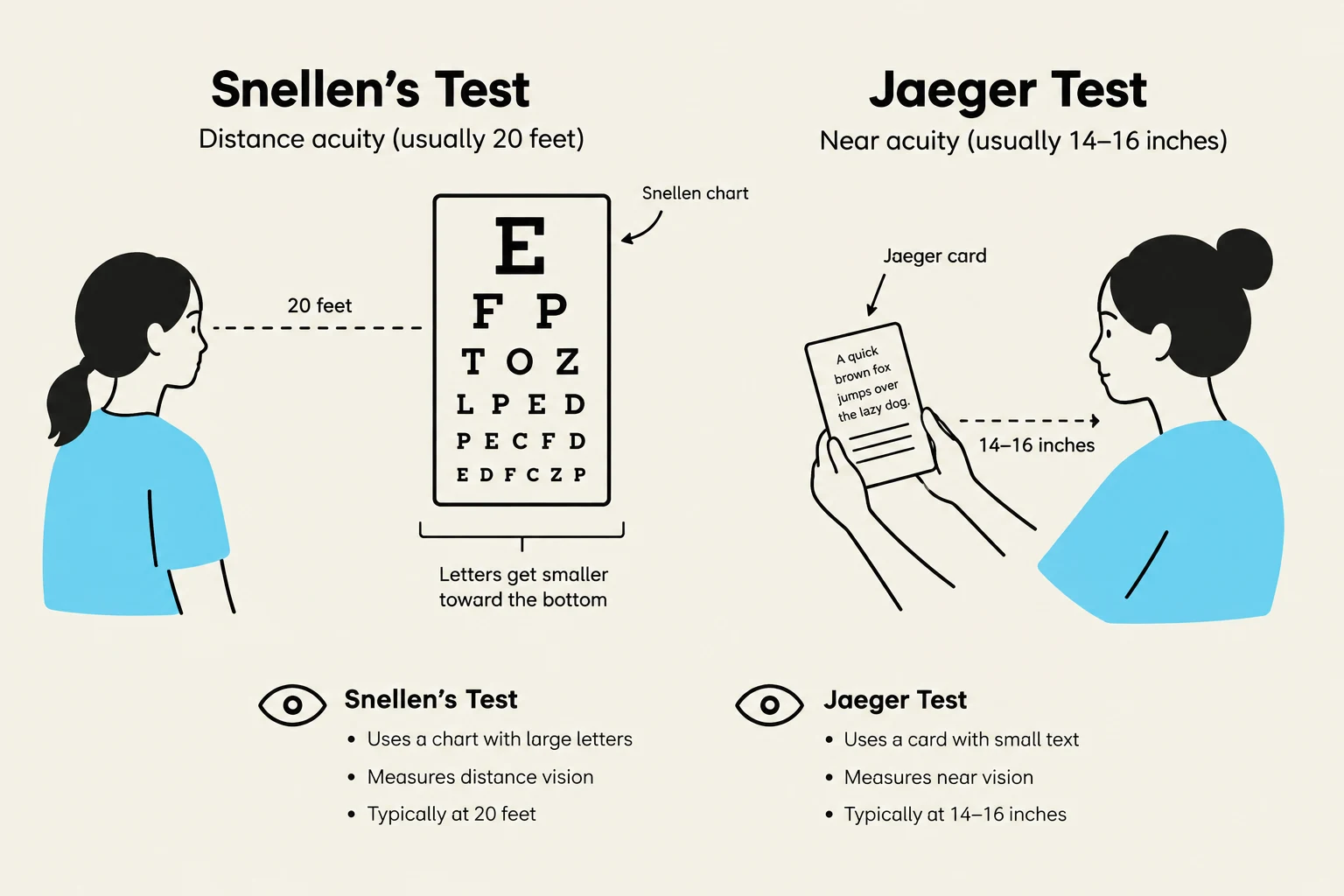
The Snellen test is the standard for assessing distance visual acuity. Patients stand at an expected distance from a chart and read the smallest line possible. The results are expressed as a fraction, such as 20/30. In this example, the numerator (20) represents the distance the patient is standing from the chart, while the denominator (30) represents the distance at which a person with normal vision could read that same line.
Jaeger Test (Near Vision)
To assess near vision, the Jaeger test is used. The patient holds a card approximately 14 inches from their eyes and is asked to read the smallest letters possible.
This illustration compares the Snellen test for distance vision and the Jaeger test for near vision, detailing their respective charts, distances, and purposes.
Pupillary Response and Cranial Nerve Assessment
The pupils provide vital information about the neurological status of the patient, particularly regarding the function of the oculomotor nerve (CN III). A normal finding is described using the acronym PERRLA: Pupils are Equal, Round, Reactive to Light, and Accommodation.
Pupillary Light Response
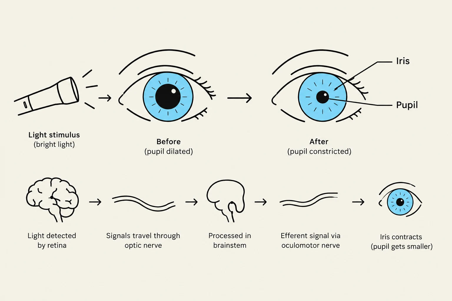
This test assesses the muscular response of the iris to light. When light is introduced, the pupils should constrict (shrink). Nurses must watch for the following abnormalities:
Anisocoria: An inequality in the size of the pupils. If this is a new finding, it is considered a medical emergency.
Miosis: Excessive constriction or smallness of the pupils, which can be caused by drugs, alcohol, stroke, or neural involvement.
Mydriasis: Excessive or prolonged dilation of the pupils, often caused by medications, drugs, or neural trauma.
Accommodation and Movement
Accommodation tests the eye's ability to focus on near objects. This is often assessed via the cover test, where the nurse observes if the eye can focus when one eye is covered, and by observing the eyes' ability to follow an object through cardinal directions.
Furthermore, the nurse must assess the extraocular muscles and movements. A key component is checking the corneal reflection; if the light reflection is symmetrical on both corneas, it is a normal finding. Asymmetry may indicate a need for further neurological assessment.
This diagram illustrates the pupillary light reflex, showing how the pupil constricts in response to bright light and dilates in dim light. It details the pathway from light detection by the retina to processing in the brainstem and the subsequent signal via the oculomotor nerve that causes the iris to contract.
Visual Field and Internal Structures
While specialized visual field testing (such as perimetry) is typically performed by optometrists, nurses may perform confrontation testing to check for gross deficits. Lesions in the brain can interrupt the messaging between the eye and the brain, resulting in visual field deficits.
When inspecting the internal ocular structures, the nurse should look at the patterns of the blood vessels. Normal vessel patterns are even, consistent, and become thicker as they approach the optic nerve. Abnormal patterns can serve as indicators for trauma, diabetes, or cardiovascular disease.
Nursing Tip: When performing an internal ocular inspection, place one hand on the patient's brow bone to stabilize yourself. Close the eye you are not using and align your right eye with the patient's right eye (and vice versa) to ensure a direct line of sight.
Summary of Cranial Nerve Involvement
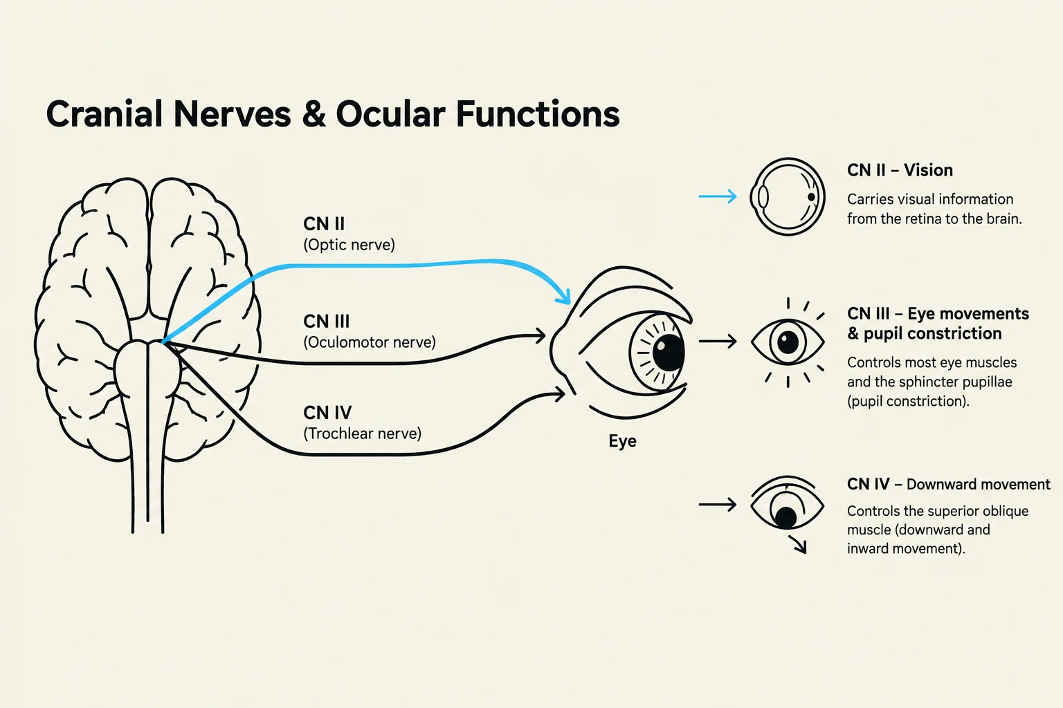
The assessment of the eyes is deeply integrated with the testing of several cranial nerves (CN):
Convergence, Cardinal directions, Pupillary reaction, Lid position
CN IV
Trochlear
Cardinal directions, Corneal reflection
CN VI
Abducens
Cardinal directions, Cover test
This diagram illustrates the cranial nerves (CN II, CN III, and CN IV) and their functions related to ocular movements and vision.
By mastering these techniques, nurses can effectively identify both structural abnormalities and neurological red flags, ensuring timely intervention for their patients.
Keep reading for free.
Create an account to finish this reading and make it part of your study routine.