An overview of normal breast anatomy, histology, and the life cycle of breast tissue, along with common benign breast changes and inflammatory conditions.
Understanding the breast requires a dual perspective: a grasp of its complex, hormone-sensitive histology and an appreciation of how it evolves across a woman's lifespan. While clinical presentations like palpable masses or nipple discharge often trigger significant patient anxiety, it is crucial to remember that only a small minority of these symptomatic lesions are actually malignant. Most breast pathologies encountered in clinical practice fall into the category of benign changes, ranging from simple inflammatory conditions to fibroepithelial neoplasms.
The Functional Unit: The Terminal Ductal-Lobular Unit (TDLU)
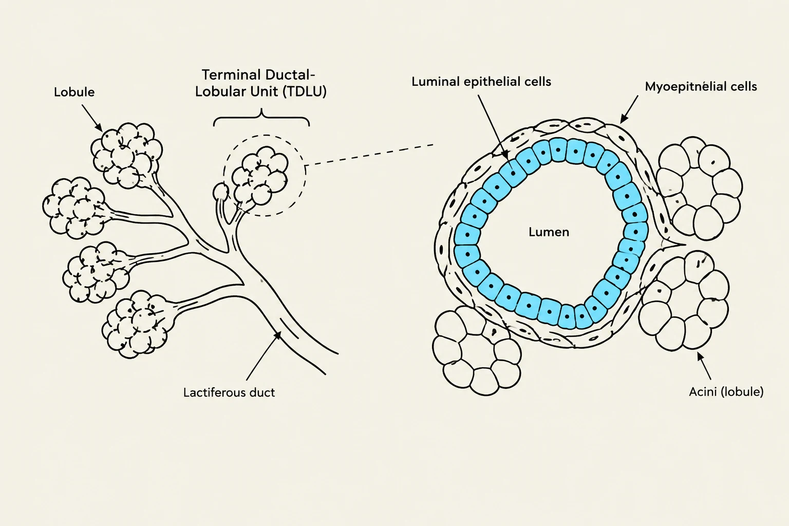
The fundamental functional unit of the breast is the Terminal Ductal-Lobular Unit (TDLU). Because the TDLU is highly sensitive to hormonal fluctuations, it serves as the primary site for both normal physiological changes and many pathological processes.
The TDLU is composed of two distinct layers of cells that line the ducts and lobules:
Luminal/Epithelial cells: These are the inner layer of cells responsible for milk production.
Basal/Myoepithelial cells: These form the outer layer, resting on the basement membrane.
Supporting these cellular structures is the stroma, which is divided into two types:
Intralobular stroma: This specialized tissue surrounds the TDLU and is highly hormone-sensitive.
Interlobular stroma: This acts as the general supportive soft tissue, similar to the connective tissue found elsewhere in the body.
Diagram illustrating the structure of a lobule, also known as a terminal ductal-lobular unit (TDLU), within the mammary gland. It shows lobules composed of acini lined by luminal epithelial cells and surrounded by myoepithelial cells, draining into a lactiferous duct.
The Life Cycle of the Breast
The anatomy of the breast is not static; it undergoes dramatic transformations driven by hormonal shifts from puberty through menopause.
Prepubertal and Reproductive Age
In the prepubertal stage, the breast consists of a rudimentary duct system that ends in terminal ductules, lacking any developed lobules. Around the onset of menarche, the ducts begin to grow and branch, lobules form, and the volume of the intralobular stroma increases as the breast reaches reproductive maturity.
Pregnancy and Lactation
The breast reaches its full functional maturity during pregnancy. As pregnancy progresses, the breasts increase in size, superficial veins become more distended, and the nipple-areolar complex becomes more pigmented. By the third trimester, the number of lobules increases significantly at the expense of the stroma.
During lactation, under the influence of prolactin, the acini become hypersecretory. Microscopically, this is characterized by the presence of lipid secretory vacuoles and the dilation of the glandular lumens.
Involution
Following menopause, the breast undergoes involution. The TDLU regresses, leading to a marked reduction in glandular tissue and a corresponding increase in fat deposition. While this process can cause ptosis (sagging), the reduction in glandular density may actually make it easier to detect cancerous lesions during imaging.
Exam Tip: When evaluating a patient's breast symptoms, always consider their stage in the life cycle. For example, a painful mass in a third-trimester pregnant woman may suggest infarction rather than a typical neoplasm.
Clinical Presentations and Inflammatory Conditions
Patients typically present with symptoms such as pain, lumpiness, palpable masses, nipple discharge, or skin changes. While many of these require a biopsy to exclude malignancy, many are benign inflammatory or structural issues.
Acute Mastitis and Periductal Mastitis
Acute mastitis is a bacterial infection (often Staphylococcus aureus or Streptococcus) that typically occurs during the first few months of breastfeeding, often entering through cracked nipple skin. It presents with redness, swelling, pain, and fever, and may require antibiotics or drainage.
In contrast, periductal mastitis (recurrent subareolar abscess) is more common in older, smoking females. Toxins from cigarette smoke cause squamous metaplasia in the large duct lining; the resulting keratin plugs the duct, leading to rupture, inflammation, and abscess formation.
Other Inflammatory and Pregnancy-Related States
Fat Necrosis: Often caused by trauma, this presents as calcifications in the stroma that can mimic carcinoma on a mammogram. Histologically, it shows foamy macrophages and multinucleate giant cells.
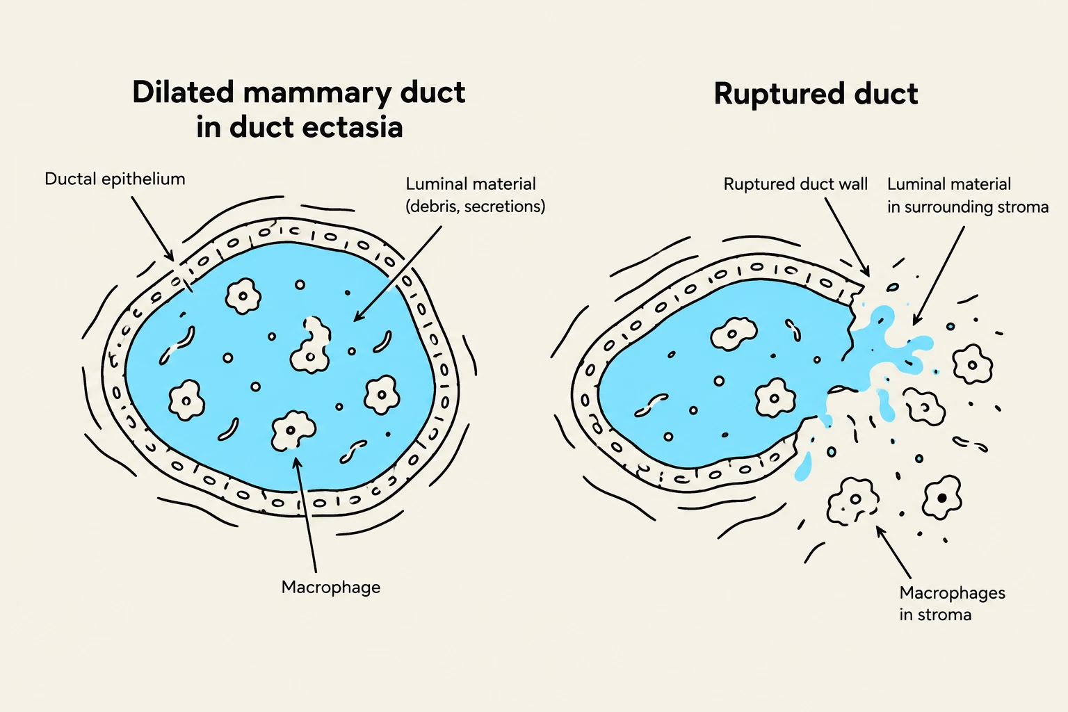
Duct Ectasia: Common in multiparous females, this involves dilated main ducts containing amorphous material and macrophages.
Infarction: Occurring most commonly in the third trimester, this happens when rapid hyperplasia causes the breast tissue to outgrow its blood supply, presenting as a painful mass.
Galactocele: This is a collection of milk in the soft tissue following the rupture of a major duct during lactation.
Diagram illustrating a dilated mammary duct in duct ectasia and a ruptured duct.
Benign Proliferative and Fibroepithelial Neoplasms
Benign changes are often categorized by whether they involve cell proliferation.
Fibrocystic Changes (Nonproliferative)
Fibrocystic changes (FCC) are the most common breast disorder and the leading cause of breast biopsies. They are considered "nonproliferative" because they do not increase the risk of cancer. FCC is characterized by:
Cystic dilatation of ducts and glands.
Stromal fibrosis.
Apocrine metaplasia.
Simple adenosis (an increase in the number of glands per lobule).
Proliferative Changes
Some benign changes involve an overgrowth of cells, which may include:
Florid Ductal Hyperplasia: An estrogen-driven overgrowth of normal cells.
Sclerosing Adenosis: Characterized by increased glands with a sclerotic intralobular stroma, often appearing as architectural distortion on imaging.
Radial Scar: A stellate or spiculated mass that features a central zone of fibroelastosis from which ducts and lobules radiate.
Intraductal Papilloma: A solitary growth within the lactiferous sinuses of the nipple; it is a common cause of spontaneous, unilateral nipple discharge.
Fibroepithelial Neoplasms
These are "biphasic" tumors, meaning they are composed of both stroma (fibro-) and glands (epithelial).
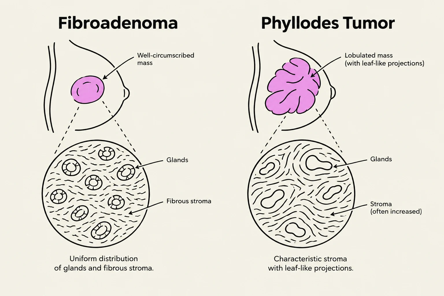
Fibroadenoma: The most common benign neoplasm in reproductive-aged women. They are typically well-defined, rubbery, and freely movable masses.
Phyllodes Tumor: These occur in older women (usually in the 6th decade) and are "stromal dominant". They are distinguished from fibroadenomas by higher stromal cellularity and a "leaf-like" appearance. While most are benign, they can grow rapidly and recur locally.
This illustration compares a fibroadenoma, characterized by a well-circumscribed mass with uniform distribution of glands and fibrous stroma, to a phyllodes tumor, which presents as a lobulated mass with leaf-like projections and increased stroma.
Gynecomastia
Finally, it is important to note gynecomastia, which is the proliferation of ducts and stroma in the male breast. This is not precancerous but is driven by a hormonal imbalance between estrogens and androgens. It can be seen during puberty, in the elderly, or in patients with cirrhosis, Klinefelter syndrome, or those using certain medications and substances like alcohol or anabolic steroids.
Summary Table: Key Benign Lesions
Condition
Key Feature
Clinical Note
Fibrocystic Change
Cysts, fibrosis, apocrine metaplasia
Most common; no increased cancer risk
Fibroadenoma
Rubbery, movable, biphasic
Most common benign neoplasm
Phyllodes Tumor
Stromal dominant, leaf-like
Occurs in older women; can be bulky
Acute Mastitis
Redness, pain, fever
Common in breastfeeding
Keep reading for free.
Create an account to finish this reading and make it part of your study routine.