DNA Library Preparation and Targeted Enrichment for Next-Generation Sequencing
This article explains the essential steps of DNA library preparation, including fragmentation, adapter ligation, and targeted enrichment techniques like hybridization capture and amplicon-based methods, crucial for Next-Generation Sequencing (NGS) in clinical diagnostics and precision medicine.
In the world of Next-Generation Sequencing (NGS), the raw genetic material extracted from a patient or a biological sample cannot be loaded directly onto a sequencing machine. The DNA is too long, too complex, and lacks the necessary "handles" for the sequencing technology to grip. To bridge this gap, scientists perform a process known as Library Preparation. This process transforms long, continuous strands of genomic DNA into a collection of short, standardized fragments called a "library."
Once the library is prepared, researchers often use Enrichment techniques to focus specifically on regions of interest, such as known cancer-causing genes. This article explores the journey of DNA from fragmentation to targeted enrichment, a workflow critical for clinical diagnostics and precision medicine.
The Foundation: DNA Fragmentation
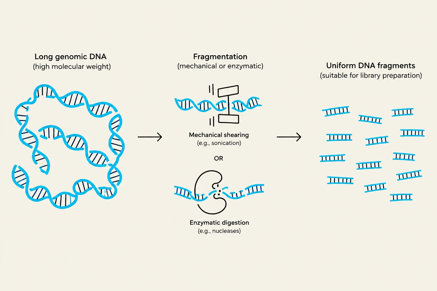
The first step in library preparation is breaking the long genomic DNA strands into smaller, manageable pieces. Most NGS platforms, such as Illumina, require DNA fragments to be within a specific size range (typically 200–500 base pairs) to ensure efficient sequencing and accurate data mapping.
There are two primary methods used to achieve this:
Physical Fragmentation: This often involves "sonication," where high-frequency sound waves create hydrodynamic shear forces that physically snap the DNA strands. This method is highly reproducible and provides a relatively uniform fragment size.
Enzymatic Fragmentation: This utilizes specific enzymes (nucleases) that cut the DNA at certain motifs or randomly. This is often gentler and can be more convenient for certain sample types, though it may introduce more bias depending on the sequence composition.
The goal is to achieve a distribution of fragment lengths that follows a predictable pattern, often represented by a bell curve.
This diagram illustrates the process of fragmenting long genomic DNA into smaller, uniform pieces suitable for library preparation, using either mechanical shearing or enzymatic digestion.
Exam Tip: If a question asks why we fragment DNA, remember that sequencing technologies have a physical limit on how much DNA they can read at once. Fragmentation ensures the fragments are small enough to be "read" by the flow cell or sensor.
Adding the Handles: Ligation of Adapters
Once the DNA is fragmented, the ends of these pieces are "messy." They may have uneven overhangs or damaged chemical groups. The next step is to repair these ends and attach adapters through a process called ligation.
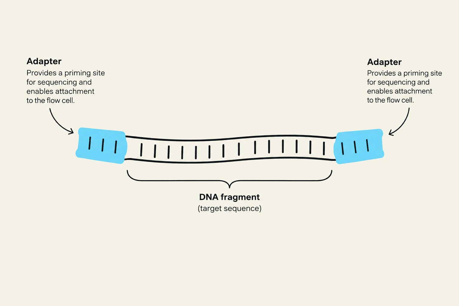
Adapters are short, synthetic, double-stranded DNA molecules that are ligated to both ends of every fragment in the library. These adapters serve several indispensable functions:
Flow Cell Attachment: They contain sequences that allow the DNA to bind to the surface of the sequencing flow cell.
Priming Sites: They provide a known starting point for the sequencing primers to bind and begin the reading process.
Indexing (Barcoding): In clinical settings, multiple patients' samples are often sequenced in a single run to save costs. Unique "index" sequences are added to the adapters, acting like digital barcodes. This allows the computer to sort the data back to the correct patient after sequencing is complete.
The ligation process involves an enzyme called DNA ligase, which catalyzes the formation of a phosphodiester bond between the DNA fragment and the adapter.
Diagram illustrating a DNA fragment with adapters attached, which are necessary for sequencing and attachment to a flow cell.
Targeted Enrichment: Focusing the Lens
Sequencing the entire genome (Whole Genome Sequencing or WGS) provides a massive amount of data, but it is expensive and often contains a lot of "noise"—information that isn't relevant to the specific clinical question being asked. For example, if a doctor wants to check for mutations in a specific set of 50 genes associated with breast cancer, sequencing the entire 3 billion base pairs of the human genome is inefficient.
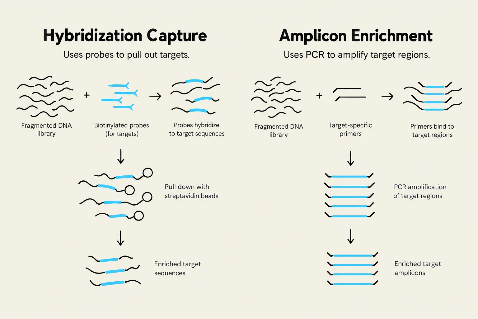
This is where Enrichment comes in. Enrichment allows us to "pull out" only the specific regions of interest from the complex library. There are two main strategies:
1. Hybridization Capture (Probe-based)
This is the most common method for large panels. Scientists design "probes"—short, single-stranded DNA sequences that are complementary to the target genes. These probes are mixed with the library. The probes find their matching sequences in the library and bind to them (hybridize). The probe-target complexes are then physically pulled out of the solution using magnetic beads.
2. Amplicon-based Enrichment (PCR-based)
This method uses specific primers to amplify only the regions of interest using Polymerase Chain Reaction (PCR). This is often faster and more efficient for very small, specific sets of genes, but it can sometimes be biased if the target regions have high GC content or complex secondary structures.
Diagram illustrating two methods for DNA enrichment: Hybridization Capture using probes and streptavidin beads, and Amplicon Enrichment using PCR and primers.
Summary of the Workflow
To visualize the entire process, consider the following mathematical relationship regarding library complexity. If N is the total number of genomic fragments and T is the number of target fragments we wish to sequence, the enrichment factor E can be thought of as:
E &= \frac{N}{T}
\end{aligned}$$
By increasing $E$ through enrichment, we ensure that the sequencing "reads" are concentrated on the most clinically relevant parts of the genome, rather than being wasted on non-coding regions.
| Step | Action | Purpose |
| --- | --- | --- |
| **Fragmentation** | Break DNA into small pieces | Make DNA compatible with sequencing hardware |
| **End Repair** | Fix DNA ends | Prepare for adapter ligation |
| **Ligation** | Attach adapters | Provide binding sites and barcodes |
| **Enrichment** | Select target regions | Increase depth of coverage for specific genes |
Through these precise steps, a raw biological sample is transformed into a highly organized, digital-ready library, enabling the powerful insights required for modern clinical genomics.
$$$
$$$
Keep reading for free.
Create an account to finish this reading and make it part of your study routine.