A detailed explanation of the neurovascular structures of the upper limb, including the brachial plexus, arteries from the aorta to the hand, venous drainage, and key anatomical spaces like the axilla, quadrangular space, and triangular interval.
The neurovascular anatomy of the upper thorax and neck involves complex networks of nerves and arteries providing motor, sensory, and nutritive support. Understanding these structures requires a systematic approach, moving from the central vessels of the thorax through the intricate branching of the brachial plexus and into the specific anatomical spaces of the axilla and arm.
The Brachial Plexus
The brachial plexus is the primary nerve network for the upper limb, originating from the ventral rami of spinal nerves $C5$ through $T1$. It is divided into two main regions: the supraclavicular portion and the infraclavicular portion.
The supraclavicular portion is found proximal to the clavicle in the posterior cervical triangle of the neck. This region contains the roots, trunks, and four specific nerves that branch directly from the ventral rami:
Dorsal scapular nerve ($C5$)
Subclavian nerve ($C5, C6$)
Suprascapular nerve ($C5, C6$)
Long thoracic nerve ($C5, C6, C7$)
The infraclavicular portion is located distal to the clavicle within the axilla, consisting of the cords and their terminal branches. Key terminal branches include the musculocutaneous, axillary, median, radial, and ulnar nerves.
Exam Tip: Note the specific dermatomes associated with these nerves. For example, $C5$ supplies the lateral arm to the proximal forearm, while $C6$ covers the lateral forearm and thumb.
Arterial Supply: From Aorta to Subclavian
Systemic blood supply to the upper limb begins at the arch of aorta. On the right side, the arch gives rise to the brachiocephalic trunk, which then divides into the right common carotid and right subclavian arteries. On the left, the left common carotid artery arises directly from the arch.
The subclavian artery is the primary trunk for the upper limb, located beneath the clavicle. It gives rise to several vital branches:
Internal thoracic artery: Runs vertically alongside the sternum.
Thyrocervical trunk: A major branch that further divides into the inferior thyroid artery and the suprascapular artery.
Dorsal scapular artery.
Diagram of the human aorta and its major branches, including the brachiocephalic artery, common carotid arteries, and subclavian arteries.
The Axillary Artery Segments
At the lateral border of the 1st rib, the subclavian artery becomes the axillary artery. This artery is clinically divided into three segments by the pectoralis minor muscle. A helpful mnemonic is that the part number equals the number of direct branches in that segment:
First part (1 branch): Located before the pectoralis minor; gives rise to the superior thoracic artery.
Second part (2 branches): Located behind the pectoralis minor; gives rise to the thoracoacromial artery and lateral thoracic artery.
Third part (3 branches): Located after the pectoralis minor; gives rise to the subscapular artery, anterior humeral circumflex artery, and posterior humeral circumflex artery.
The subscapular artery is particularly significant as it branches into the circumflex scapular artery (traveling around the scapula) and the thoracodorsal artery (traveling toward the latissimus dorsi).
The Brachial Artery and Venous Drainage
Once the axillary artery passes the inferior border of the teres major muscle, it is renamed the brachial artery (or the deep artery of the arm). The brachial artery provides critical supply to the arm via the profunda brachii artery (which travels deep) and the superior and inferior ulnar collateral arteries. Eventually, the brachial artery bifurcates into the radial and ulnar arteries.
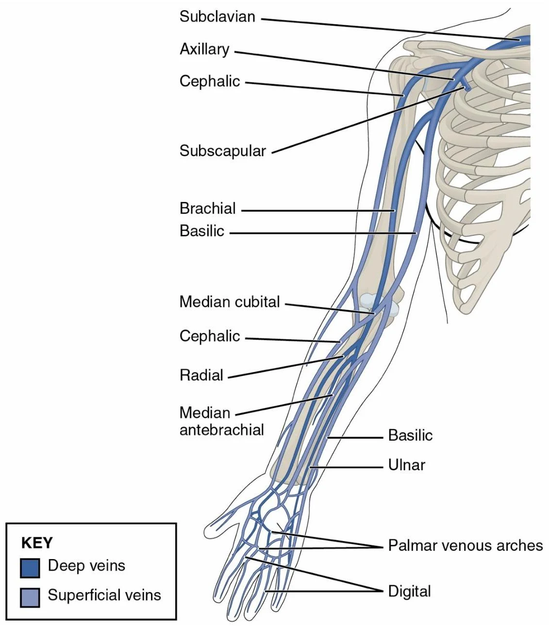
For venous return, the arm utilizes both deep and superficial systems. Key veins include the cephalic vein, the brachial vein, and the medial cubital vein.
Diagram illustrating the superficial and deep veins of the human arm, including labels for major veins such as the subclavian, axillary, cephalic, brachial, basilic, radial, and ulnar veins, as well as the palmar venous arches and digital veins.
Anatomical Spaces: Axilla, Quadrangular Space, and Triangular Interval
The axilla is a pyramidal space that serves as a passageway for neurovascular structures. Its boundaries are:
Medial: Serratus anterior
Anterior: Pectoralis major and minor
Posterior: Subscapularis, teres major, and latissimus dorsi
Lateral: Humerus
Within the posterior aspect of the shoulder and axilla, there are specific "windows" or spaces formed by muscles that allow for the passage of nerves and vessels:
Quadrangular Space
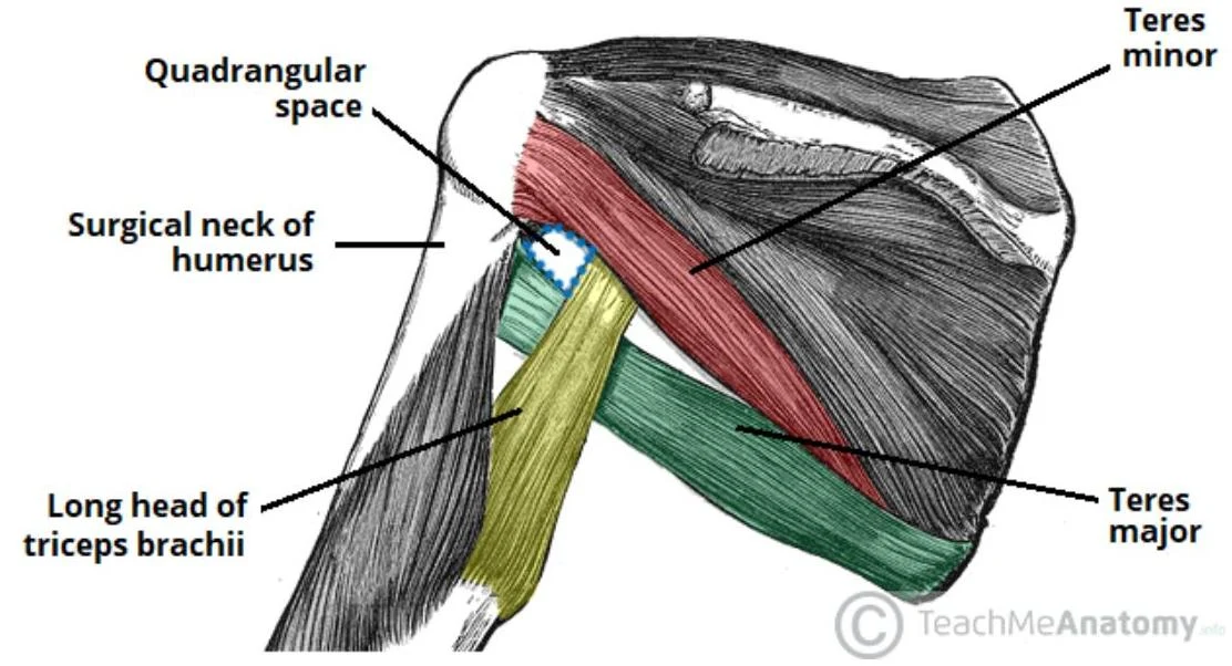
This space is bounded by the teres major, teres minor, humerus, and the long head of the triceps brachii. It is a critical conduit for the axillary nerve and the posterior humeral circumflex artery.
An anatomical illustration showing the muscles and spaces of the posterior shoulder and upper arm, including the quadrangular space, surgical neck of the humerus, teres minor, teres major, and the long head of the triceps brachii.
Triangular Interval
Located inferior to the quadrangular space, this interval is bounded by the teres major, the long head of the triceps brachii, and the lateral head of the triceps brachii. It contains the profunda brachii artery and the radial nerve.
Anatomical illustration of the borders of the triceps brachii muscle, including the lateral head, long head, and the superior border indicated by the teres major muscle.
Clinical Correlation: Damage to the structures within these spaces can lead to specific deficits. For example, injury to the axillary nerve in the quadrangular space can result in deltoid muscle weakness and loss of sensation over the lateral shoulder.
Keep reading for free.
Create an account to finish this reading and make it part of your study routine.