This content explains the two major classes of neurotransmitter receptors: ionotropic and metabotropic. It details their mechanisms of action, including G-protein signaling and second messenger cascades, and discusses pharmacological analysis methods like EC50 and IC50 for measuring receptor potency.
The orderly function of the human brain relies on a complex series of chemical reactions, most notably those occurring during synaptic transmission. When a neuron communicates with its target, it releases neurotransmitters that travel across the synaptic cleft to bind with specific proteins on the post-synaptic membrane known as receptors. These receptors act as the "interpreters" of the chemical signal, converting a molecular binding event into a functional change in the cell.
The Two Major Receptor Classes
Neurotransmitters do not all act in the same way. Instead, they bind to different receptor subtypes that dictate the speed and nature of the cellular response. These subtypes are broadly categorized into two functional classes: ionotropic and metabotropic receptors.
Ionotropic Receptors: Fast and Direct
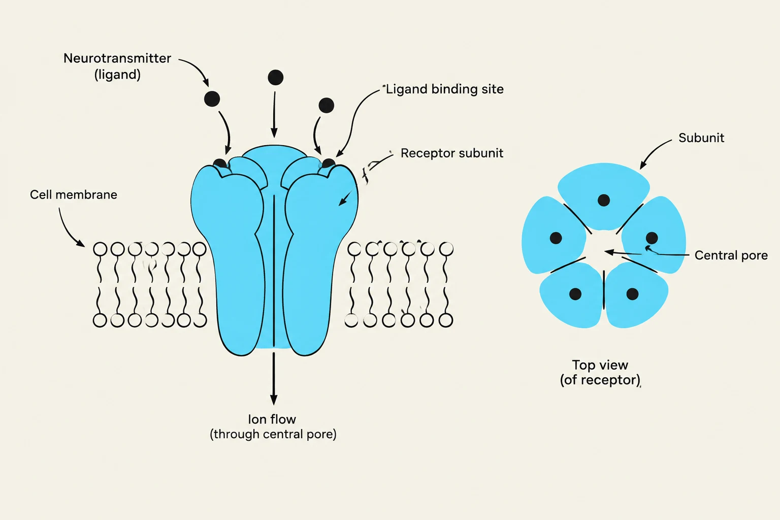
Ionotropic receptors are ligand-activated ion channels. These are large protein complexes composed of multiple membrane-spanning domains called subunits. These subunits organize to form a central pore that allows specific ions to flow into or out of the cell.
The structural hallmark of these receptors is the presence of ligand binding sites located on one or more of the subunits. The number of occupied binding sites often correlates with the duration of the channel opening; the more transmitter molecules bound, the longer the channel remains open. Furthermore, the amino acids lining the pore act as a "selectivity filter," ensuring that only specific ions (such as Na+, $K^+$, or $Cl^-$) can pass through.
Diagram illustrating a neurotransmitter binding to a receptor on the cell membrane, leading to ion flow through a central pore. A top view of the receptor shows its subunit structure.
Metabotropic Receptors: Slow and Modulatory
Metabotropic receptors, also known as G-protein-coupled receptors (GPCRs), operate through a more complex, indirect mechanism. Unlike the multi-subunit channels of ionotropic receptors, most metabotropic receptors consist of a single polypeptide chain that weaves through the membrane seven times (seven membrane-spanning alpha helices).
The function of a metabotropic receptor is split between its two sides:
Extracellular loops: These form the binding sites for the neurotransmitter.
Intracellular loops: These interact with G-proteins, the molecular switches that initiate the cellular response.
Exam Tip: If a question asks about the primary difference in speed between receptor types, remember that ionotropic receptors are "fast" (direct ion flow), while metabotropic receptors are "slow" (requiring chemical intermediaries).
G-Protein Signaling and Second Messenger Cascades
The hallmark of metabotropic signaling is the involvement of G-proteins. A G-protein is a heterotrimeric complex consisting of three subunits: α (alpha), β (beta), and γ (gamma).
The G-Protein Cycle
In its inactive state, the α subunit is bound to GDP (guanosine diphosphate) and the three subunits "float" together in the membrane. The activation process follows these steps:
Activation: An activated receptor bumps into the G-protein, causing the α subunit to exchange its bound GDP for GTP (guanosine triphosphate).
Dissociation: The GTP-bound α subunit splits from the βγ complex.
Effector Interaction: Both the Gα-GTP and the Gβγ subunits can now interact with other proteins to produce effects.
Inactivation: The Gα subunit eventually converts its GTP back into GDP, causing it to recombine with Gβγ, resetting the cycle.
The Shortcut vs. The Cascade
G-proteins can exert their effects through two distinct routes:
The Shortcut Pathway: The Gβγ subunits can bind directly to nearby ion channels, opening them with relatively fast response times (approx. 30ms).
The Second Messenger Cascade: The Gα subunit can activate enzymes, which then produce intracellular signaling molecules called "second messengers".
Diagram illustrating the G-protein activation cycle, showing the steps from an inactive state to signal transmission.
Common cascades include:
Adenylyl Cyclase Pathway:Gαs stimulates adenylyl cyclase to produce cAMP, which activates Protein Kinase A (PKA). Conversely, Gαi inhibits this process.
Phospholipase C Pathway:Gαq activates phospholipase C, leading to the production of second messengers that activate Protein Kinase C (PKC) and increase intracellular Ca2+.
These kinases then phosphorylate target proteins, such as ion channels or transcription factors, to alter their activity.
Signal Amplification and Pharmacological Analysis
One of the greatest evolutionary advantages of the metabotropic system is signal amplification. Because one activated receptor can trigger multiple G-proteins, and each G-protein can activate enzymes that produce thousands of second messenger molecules, a tiny amount of neurotransmitter can lead to a massive cellular response.
Measuring Receptor Potency
Pharmacologists study these systems by applying drugs to measure postsynaptic responses. They use two main types of compounds:
Agonists: Chemicals that mimic the natural neurotransmitter.
Antagonists: Chemicals that block the receptor's activity.
To quantify how well these drugs work, we use two key metrics: EC50 and IC50.
Metric
Definition
Context
EC50
The concentration of an agonist that produces 50% of its maximal effect.
Measures potency/affinity of stimulators.
IC50
The concentration of an antagonist that reduces the agonist response by 50%.
Measures potency/affinity of blockers.
Note on Affinity: For both agonists and antagonists, a lower numerical value for EC50 or IC50 indicates a higher potency (affinity), because it takes less of the drug to achieve the half-maximal effect.
Diversity of Subtypes
Pharmacological analysis reveals that a single neurotransmitter can act on many different receptor subtypes, which may be located on the same neuron or distributed differently across the body. For example, acetylcholine acts on nicotinic receptors (ionotropic) in the peripheral nervous system, but on muscarinic receptors (metabotropic) in the central nervous system. Furthermore, the diversity of receptor function is driven by the complex assembly of subunits; for instance, $GABA_A$ receptors can combine different polypeptide subunits to create a vast array of functional variations.
Keep reading for free.
Create an account to finish this reading and make it part of your study routine.