Explores the sensory system's role in interfacing with the environment, distinguishing sensation from perception, and detailing sensory transduction, receptor types, adaptation, and coding mechanisms. Covers somatosensory pathways and the physiology of pain.
The sensory system serves as the body's primary interface with the world, constantly monitoring both the external environment and internal physiological states. By detecting changes and transmitting this information to the Central Nervous System (CNS), the sensory system allows for complex integration, enabling organisms to respond to threats, seek nourishment, and maintain homeostasis.
Sensation vs. Perception
To understand sensory neurophysiology, one must distinguish between the raw data received by the body and the conscious experience of that data.
Sensation is the initial process of detecting a stimulus via sensory receptors. It is the physiological detection of energy, such as a change in temperature or a mechanical pressure.
Perception, conversely, is a higher-order process. It involves the organization, interpretation, and conscious awareness of those sensations. For example, while the sensation of pain might be the detection of tissue damage, the perception is the subjective experience of an "achy joint". It is important to note that perception is not always an accurate reflection of reality; optical illusions and perceptual gaps demonstrate that the brain often fills in missing information or misinterprets sensory input.
Sensory input is generally categorized into two types:
Somatic Sensations: Derived from the skin, muscles, bones, and tendons (e.g., touch, pressure, temperature, and pain).
Visceral Sensations: Derived from internal organs, such as the heart, lungs, or GI tract, often involving pressure, stretch, or chemical changes.
Sensory Transduction and Receptor Types
The core mechanism of any sensory system is sensory transduction: the conversion of a physical or chemical stimulus into a biological signal called a receptor potential. A receptor potential is a graded potential that changes in magnitude based on the stimulus.
Every stimulus possesses a specific form of energy known as its modality (e.g., light waves, chemical concentrations, or mechanical force). Each sensory receptor is tuned to an adequate stimulus, which is the specific modality it responds to most efficiently. While a receptor might respond to a non-adequate stimulus if the intensity is extremely high, its primary function is tied to its adequate modality.
Based on their adequate stimuli, receptors are classified into five major divisions:
Mechanoreceptors: Respond to mechanical force such as touch, pressure, or vibration.
Chemoreceptors: Respond to chemical concentrations.
Thermoreceptors: Respond to temperature changes.
Nociceptors: Respond to pain (noxious stimuli).
Photoreceptors: Respond to light.
Exam Tip: Do not confuse a sensory receptor with a hormone or neurotransmitter receptor. A sensory receptor is the distal end of an afferent neuron (or a structure associated with it) that detects environmental energy, whereas a hormone receptor is a protein that binds a chemical ligand.
Adaptation: Phasic vs. Tonic Receptors
The magnitude of a receptor potential is influenced by the stimulus strength, the rate of change in that strength, and temporal summation. Over time, many receptors undergo adaptation, which is a decrease in the magnitude of the receptor potential even if the stimulus remains constant.
Receptors are categorized by their rate of adaptation:
Rapidly Adapting (Phasic) Receptors: These cease responding quickly to a constant stimulus. They are ideal for monitoring movement or rapid changes, such as vibrations or the initial sensation of a smell.
Slowly Adapting (Tonic) Receptors: These maintain a persistent potential for the duration of the stimulus. They are essential for signaling the intensity of prolonged stimuli, such as maintaining awareness of body posture and joint position.
This diagram illustrates the difference in receptor potential and afferent neuron action potentials between rapidly adapting receptors (top) and slowly adapting receptors (bottom) in response to a stimulus. The top graph shows a brief burst of action potentials for vibration and pressure, while the bottom graph shows sustained action potentials for joint and muscle movement and posture.
Sensory Pathways and Coding
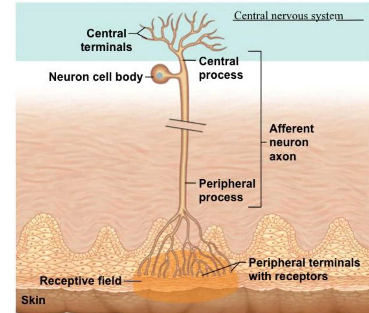
Sensory information travels through organized neural pathways. A sensory unit consists of a single afferent neuron and all the sensory receptors connected to it. The specific area of the body that activates a particular sensory unit is called its receptive field.
Diagram illustrating a sensory neuron with its cell body, central and peripheral processes, and terminals with receptors in the skin, part of the central nervous system.
The CNS uses "Labeled Lines" to interpret these signals, meaning specific pathways transmit specific modalities to specific regions of the brain. This involves a serial process of three neurons:
First-order neuron: The afferent neuron carrying the signal from the receptor.
Second-order neuron: An interneuron in the CNS that transmits info to the thalamus.
Third-order neuron: Transmits information from the thalamus to the proper region of the cerebral cortex.
Diagram illustrating the pathway of a sensory neuron from receptors to the cortex, passing through the spinal cord/brainstem and thalamus.
Sensory Coding Mechanisms
The nervous system must "code" the stimulus into electrical signals. It does this through three primary methods:
Modality Coding: Determined by which specific receptor is activated.
Intensity Coding: Coded via frequency coding (increasing the frequency of action potentials in a single neuron) and population coding (recruiting more sensory receptors and adjacent sensory units).
Location Coding: Determined by the size and number of receptive fields and the process of lateral inhibition.
Lateral Inhibition is a crucial mechanism where a strongly activated afferent neuron causes presynaptic inhibition of adjacent sensory units. This enhances the contrast between the center of a stimulus and its periphery, allowing the brain to pinpoint the exact location of a stimulus with high acuity.
Somatosensory Pathways: Anterolateral vs. Dorsal Column
The somatosensory system utilizes two distinct ascending pathways to transmit information to the cortex:
Feature
Anterolateral (Spinothalamic) Pathway
Dorsal Column Pathway
Primary Modalities
Pain and Temperature
Proprioception, Fine touch, Vibration
Synapse (1st to 2nd order)
Occurs in the spinal cord gray matter
Occurs in the brainstem
Decussation (Crossing)
2nd order neuron crosses in spinal cord
2nd order neuron crosses in brainstem
The Physiology of Pain (Nociception)
Pain is not just a sensory modality but a complex experience designed as an evolutionary adaptation to protect against tissue damage.
Nociception occurs when noxious stimuli (heat, intense mechanical force, or chemicals like prostaglandins) activate free nerve endings. Pain can be classified into several mechanisms:
Nociceptive: Caused by actual tissue damage or inflammation.
Neuropathic: Resulting from injury to the neural structures themselves (e.g., stroke or trauma).
Psychogenic: Caused or prolonged by mental or emotional factors.
A unique phenomenon in pain is referred pain. This occurs because visceral and somatic afferents often converge onto the same second-order neurons. Consequently, the brain may misinterpret a signal from an internal organ as coming from a somatic area (like the skin).
Diagrams illustrating the sensory pathway of pain signals from the skin to the spinal cord and brain, and a diagram showing referred pain patterns associated with internal organs.
Clinical Note:Hyperalgesia refers to an increased sensitivity to painful stimuli, where pain may persist for hours after the original stimulus has been removed.
Pain Management
Pain management strategies target different parts of the pathway:
NSAIDs (e.g., Ibuprofen): Inhibit prostaglandin synthesis at the site of injury.
Opioids (e.g., Morphine): Act within the CNS to alter the perception of pain.
Local Anesthetics: Block the transmission of the noxious stimulus along the nerve.
TENS (Transcutaneous Electrical Nerve Stimulation): Uses electrical stimulation to activate touch receptors, which then use lateral inhibition to "turn off" smaller pain afferents.
Keep reading for free.
Create an account to finish this reading and make it part of your study routine.