Neurophysiology of Neuronal Signaling: Local and Action Potentials
Explore the fundamental principles of neurophysiology, including the resting membrane potential, local potentials (depolarization and hyperpolarization), and the all-or-none action potential. Learn about the mechanisms of signal propagation, neurotransmission, and clinical correlations.
Neurophysiology is the study of how neurons generate and transmit electrical signals to communicate information throughout the body. These signals are not random; they are driven by the electrochemical gradient, which is the interplay between a concentration gradient (ions moving from high to low concentration) and an electrical gradient (ions moving toward opposite charges). To understand how a neuron "fires," we must first understand the state of the cell when it is at rest.
The Resting Membrane Potential
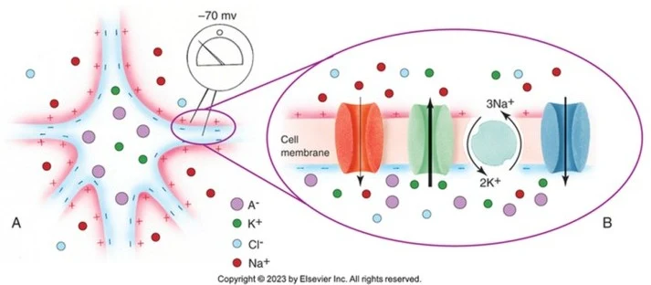
The resting membrane potential is a steady-state condition where the interior of the neuron is negatively charged relative to the exterior, typically ranging from −60 mV to −70 mV. This negative charge is essential for creating the potential energy required for signaling.
This state is maintained by three primary mechanisms:
Leak Channels: Passive diffusion occurs through channels that are always open. Most of these are K+ leak channels, which allow K+ to exit the cell.
The Na+/K+ Pump: This active transport mechanism uses energy from ATP to move 3 Na+ ions out of the cell for every 2 K+ ions moved in. This maintains the concentration imbalance necessary for the gradient.
Anions: Large, negatively charged molecules, such as proteins and phosphates, are trapped inside the cell because they are too large to diffuse through the channels.
Diagram illustrating the resting membrane potential of a neuron, showing ion concentrations and transport mechanisms across the cell membrane.
Local Potentials: Graded Changes
When a neuron is stimulated, the membrane potential changes. These changes are categorized as either depolarization (the membrane becomes less negative/more positive) or hyperpolarization (the membrane becomes more negative).
Local potentials are graded changes in membrane potential that spread only short distances via passive propagation. They vary in both amplitude and duration and can be either excitatory or inhibitory.
Types of Local Potentials
Receptor Potentials: These are generated at the peripheral receptors of a sensory neuron when they are exposed to stimuli like stretching, compression, or thermal changes. For example, stretching a muscle can open Na+ or Ca2+ channels, generating a receptor potential.
Synaptic Potentials: These occur at the postsynaptic membrane when neurotransmitters bind to ligand-gated channels.
Excitatory Postsynaptic Potentials (EPSPs): These cause depolarization, making the membrane potential more positive than the resting state.
Inhibitory Postsynaptic Potentials (IPSPs): These cause hyperpolarization, making the membrane potential more negative.
Summation
Because local potentials are small, they must often be integrated to reach the threshold required for an action potential. This occurs through:
Temporal Summation: A single presynaptic neuron fires a series of small potentials in rapid succession (within milliseconds).
Spatial Summation: Multiple different presynaptic neurons fire simultaneously at different locations on the same postsynaptic neuron.
Action Potentials: The "All-or-None" Signal
While local potentials are passive and short-lived, the action potential is an active, large-scale depolarization that is repeatedly regenerated to transmit information over long distances. An action potential is "all-or-none," meaning that once a stimulus reaches the threshold potential (approximately −55 mV), an action potential will fire with a constant amplitude regardless of the stimulus strength.
This diagram illustrates the process of action potential generation in a neuron, detailing the changes in membrane potential and ion channel activity during resting potential, depolarization, repolarization, and hyperpolarization.
The phases of an action potential include:
Depolarization: Upon reaching threshold, voltage-gated Na+ channels open rapidly, allowing an influx of Na+ that makes the interior of the cell positive.
Repolarization: Na+ channels inactivate, and voltage-gated K+ channels open, allowing K+ to exit the cell and restore a negative charge.
Hyperpolarization: K+ channels often remain open longer than necessary, causing the membrane potential to drop below the normal resting level.
Refractory Periods and Propagation
Following an action potential, the neuron enters a refractory period. During the absolute refractory period, it is impossible to trigger another action potential because the Na+ channels are inactivated. During the relative refractory period, a second action potential can be triggered, but it requires a much stronger stimulus because the cell is hyperpolarized.
To ensure rapid communication, neurons use structural adaptations:
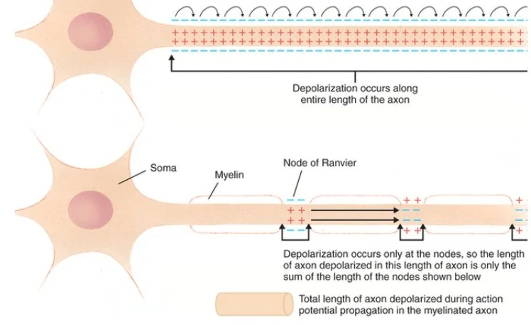
Myelination: Glial cells (Oligodendrocytes in the CNS and Schwann cells in the PNS) wrap axons in insulating myelin.
Saltatory Conduction: Instead of continuous depolarization, the signal "jumps" between the nodes of Ranvier (gaps in the myelin), significantly increasing conduction velocity.
Diagram illustrating the process of depolarization along a myelinated axon, highlighting the difference between unmyelinated and myelinated nerve impulse propagation.
Neural Communication: Neurotransmitters and Neuromodulators
Information is passed between neurons at the synapse, which consists of the presynaptic terminal, the synaptic cleft, and the postsynaptic terminal.
Neurotransmitters (Fast-Acting)
Neurotransmitters carry chemical signals across the synapse to induce rapid changes in membrane potential.
Ionotropic (Fast-acting): These bind directly to ligand-gated ion channels. The receptor and the channel are a single functional unit, allowing for millisecond-scale responses.
Glutamate: The primary fast excitatory neurotransmitter in the CNS.
GABA: The major inhibitory neurotransmitter in the CNS.
Acetylcholine (ACh): Essential for muscle contraction at the neuromuscular junction.
Neuromodulators (Slow-Acting)
Neuromodulators are a subset of chemicals released into extra-synaptic sites (outside the synapse). They do not cause rapid signal transmission but instead alter the properties of ion channels to adjust the activity of many neurons simultaneously.
Metabotropic (Slow-acting): These bind to G-protein coupled receptors (GPCRs), which activate a second messenger system. This can lead to long-term changes, such as altering gene expression or increasing the number of receptors on a dendrite.
Examples: Dopamine (linked to Parkinson's), Serotonin (mood regulation), and Endorphins (pain modulation).
💡 Exam Tip: Agonists vs. Antagonists
Agonists (e.g., Levodopa for Parkinson's) mimic the effects of naturally occurring neurotransmitters by binding to and activating receptors.
Antagonists (e.g., Botulinum Toxin) impede the effects of neurotransmitters by either blocking the release of the chemical or preventing it from binding to the receptor.
Clinical Correlations
Demyelinating Diseases
When the insulating myelin is destroyed, signal conduction is disrupted, leading to weakness, loss of coordination, and sensory deficits.
Guillain-Barré Syndrome: An autoimmune attack on the myelin of the Peripheral Nervous System (PNS), often following an infection.
Multiple Sclerosis (MS): An autoimmune attack on the oligodendrocytes in the Central Nervous System (CNS), causing plaques in the white matter.
Neuromuscular Junction Disorders
Lambert-Eaton Myasthenic Syndrome (LEMS): Antibodies attack the voltage-gated Ca2+ channels in the presynaptic terminal, reducing the release of Acetylcholine.
Myasthenia Gravis (MG): Antibodies attack and destroy the nicotinic ACh receptors on the postsynaptic muscle membrane, leading to muscle weakness that worsens with repetitive use.
🔍 Clinical Case Study: Myasthenia Gravis
A patient presents with ptosis (drooping eyelids) that worsens in the evening and improves with rest. This is a hallmark of Myasthenia Gravis because repetitive use of the muscle depletes the available receptors, leading to increased weakness as the day progresses.
Keep reading for free.
Create an account to finish this reading and make it part of your study routine.