An overview of the nervous system's cellular components, detailing the morphology and function of neurons and the essential support roles of glial cells in both the Central and Peripheral Nervous Systems.
The nervous system is a marvel of biological engineering, functioning through a complex interplay of specialized cells that transmit, integrate, and process information. At the heart of this system are neurons—the functional units responsible for electrochemical communication—and glia, the essential support cells that maintain the physiological environment necessary for neuronal survival. To understand how the brain and spinal cord control everything from a heartbeat to a complex thought, one must first master the diverse morphologies of neurons, the intricate internal machinery of the cell, and the distinct roles played by glial cells in both the Central Nervous System (CNS) and the Peripheral Nervous System (PNS).
The Morphological Classes of Neurons
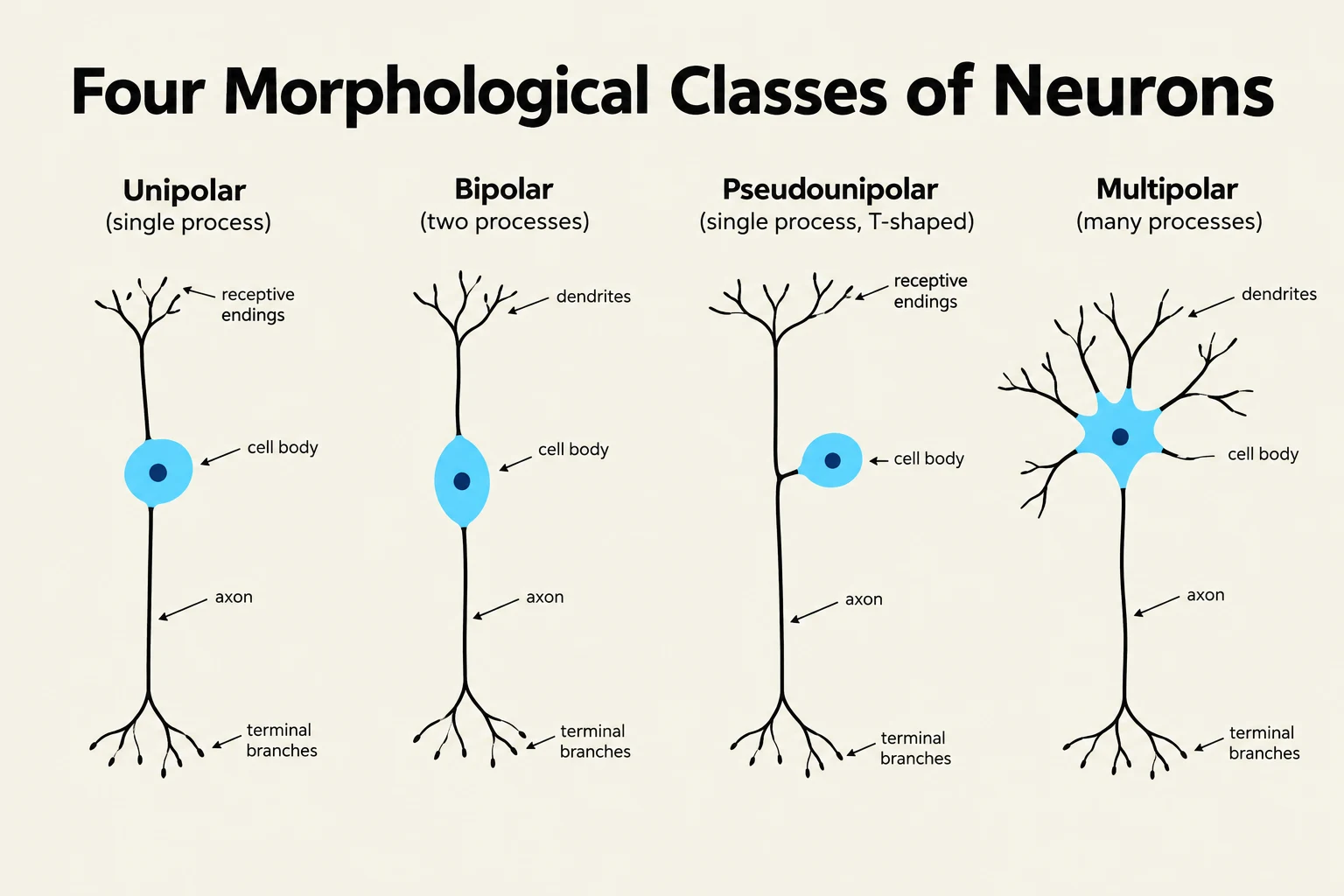
While all neurons share the fundamental goal of signal transmission, their physical shapes, or phenotypes, vary widely to suit their specific functional roles. We categorize neurons into four primary morphological classes based on the number and arrangement of their processes (axons and dendrites).
Unipolar Neurons
The unipolar neuron is the simplest morphological form. Unlike most vertebrate neurons, it lacks true dendrites; instead, the cell body itself serves to receive and integrate incoming information. While rare in the vertebrate nervous system, these are commonly observed in invertebrate nervous systems.
Bipolar Neurons
Bipolar neurons possess two distinct processes extending from opposite poles of the cell body: one acting as a dendrite-like receiver and the other as an axon-like transmitter. This specialized structure is highly efficient for relaying sensory information along a direct path, and these neurons are commonly found in sensory organs such as the retina.
Pseudounipolar Neurons
A pseudounipolar neuron is essentially a specialized subtype of the bipolar neuron. During embryonic development, two processes emerge from the cell body but eventually fuse into a single process that subsequently bifurcates (splits) away from the soma. This structure is characteristic of somatic sensory neurons, which are responsible for carrying information regarding touch, pain, temperature, and proprioception (the sense of body position) from the periphery to the CNS.
Multipolar Neurons
The most prevalent neuron type in the human brain and spinal cord is the multipolar neuron. These cells feature a highly complex dendritic array—allowing them to receive vast amounts of integrated input—and a single, often branching, axon for signal output.
Student Tip: Don't confuse morphology with function! While a neuron's shape often hints at its job (e.g., bipolar in the retina), remember that "multipolar" refers to the structure (many dendrites), not necessarily the complexity of the thought it processes.
Diagram illustrating the four morphological classes of neurons: unipolar, bipolar, pseudounipolar, and multipolar, with labels for their key components such as cell body, axon, dendrites, and terminal branches.
Neurocytology: The Internal Machinery
A neuron is not merely a wire; it is a metabolically active cell requiring constant maintenance. This internal machinery, known as neurocytology, can be divided into the genetic control center, the endomembrane system, and the structural cytoskeleton.
The Nucleus and the Endomembrane System
The nucleus serves as the repository of DNA, directing all essential processes including gene expression, protein synthesis, cellular growth, and long-term maintenance.
To translate genetic instructions into functional proteins, the neuron utilizes the endomembrane system, an interconnected network of membranes.
Rough Endoplasmic Reticulum (ER): This organelle is studded with ribosomes and is responsible for building proteins that are destined for secretion or for integration into the cell membrane.
Golgi Apparatus: Once proteins are synthesized in the ER, they are sent to the Golgi apparatus, which acts as a "packaging center," sorting and placing proteins into vesicles for their final cellular destinations.
Clinical Note: Errors in this system are not just theoretical. Misfolded proteins within the endomembrane system are implicated in severe neurodegenerative conditions, such as prion disorders.
The Cytoskeleton and Axonal Transport
Because neurons can be incredibly long (some axons extending from the spine to the toes), they require a robust internal scaffolding to maintain shape and move materials. This is provided by three types of filaments:
Microtubules: These act as the primary structural scaffolds and serve as the "tracks" for intracellular transport.
Neurofilaments: These provide longitudinal support and are critical for regulating the diameter of the axon.
Microfilaments (Actin): These are involved in anchoring receptors, ion channels, and the machinery required for presynaptic release.
The movement of materials along these microtubule tracks is a highly organized process called axonal transport.
Anterograde Transport: Driven by the motor protein kinesin, this mechanism moves vesicles and materials away from the cell body (soma) toward the axon terminal.
Retrograde Transport: Driven by the motor protein dynein, this mechanism moves materials from the axon terminal back toward the cell body.
Glial Cells: The Essential Support Network
For a long time, glial cells were thought to be mere "glue" holding neurons together. We now know they are at least as numerous as neurons and are indispensable for metabolic and physiological function. Glia are categorized based on their location in either the Central Nervous System (CNS) or the Peripheral Nervous System (PNS).
Central Nervous System (CNS) Glia
The CNS, comprising the brain and spinal cord, relies on several specialized glial types:
Astrocytes: These are the "managers" of the CNS. They act as structural scaffolds for migrating neurons during development, monitor synaptic activity to provide chemical feedback, and maintain the blood-brain barrier (BBB) via their specialized "endfeet". The BBB is formed by endothelial tight junctions that restrict the movement of large or harmful molecules into brain tissue.
Oligodendrocytes: These cells are responsible for myelination within the CNS. A single oligodendrocyte is highly efficient, capable of providing myelin sheaths to dozens of different axons.
Microglia: Acting as the resident immune system of the brain, microglia are phagocytic "scavengers" that respond rapidly to injury or infection. While vital for healing, they can sometimes contribute to chronic issues like neuropathic pain if they remain overactive.
Ependymal Cells: These epithelial-like cells line the ventricles of the brain and the central canal of the spinal cord, regulating the chemical flow of cerebrospinal fluid (CSF) into brain tissue.
Peripheral Nervous System (PNS) Glia
The PNS, which includes all nerves and ganglia outside the CNS, utilizes a different set of support cells:
Schwann Cells: These are the PNS counterparts to oligodendrocytes. However, there is a key difference: while one oligodendrocyte can support many axons, a single myelinating Schwann cell supplies only one axon. Crucially, Schwann cells play a major role in nerve repair, proliferating after injury to stimulate axon elongation and promote regeneration.
Satellite Glial Cells: Located within sensory, sympathetic, and parasympathetic ganglia, these cells closely enclose individual neuron cell bodies. They maintain a tight physical connection (often only 20nm apart) and communicate with neurons via the release of chemicals like ATP, GABA, and glutamate to regulate the local microenvironment.
Diagrams illustrating oligodendrocytes in the CNS and Schwann cells in the PNS, highlighting their roles in myelinating axons.
Clinical Application: Why Distinctions Matter
Understanding the difference between neuronal components and glial support is not just an academic exercise; it is clinically vital.
Consider the case of Myasthenia Gravis. In this autoimmune disease, the body produces antibodies that attack acetylcholine receptors at the neuromuscular junction. This is a disease of the synapse (the communication point between a neuron and a muscle), specifically targeting a protein on the neuronal/muscle membrane. It is not a disease of the glial cells (like demyelinating diseases such as Multiple Sclerosis).
By distinguishing whether a pathology targets the neuron's signaling machinery (like receptors) or the glial support structures (like myelin), clinicians can determine whether to treat the neurotransmission process or the protective insulation of the nerve.
Exam Check: If a question asks which cell type is responsible for the blood-brain barrier, look for Astrocytes. If it asks which cell type promotes regeneration in the peripheral nerves, the answer is Schwann cells.
Keep reading for free.
Create an account to finish this reading and make it part of your study routine.