Neuronal Action Potentials and Synaptic Communication
This study sheet covers the transmission of nerve impulses by neurotransmitters, the properties of neuronal action potentials, and their role in nervous system functions like learning and memory.
The nervous system is a marvel of biological engineering, designed to achieve two seemingly contradictory goals: extreme speed for survival reflexes and immense flexibility for complex thought. This balance is made possible by the precise electrochemical language of neurons. By understanding how an electrical impulse travels down an axon and how that signal is translated into a chemical message at a synapse, we can begin to understand the very foundations of learning, memory, and motor control.
The Electrical Impulse: Action Potentials
An action potential is a rapid, temporary change in the electrical membrane potential of a neuron. This process is governed by the movement of ions across the cell membrane through specialized protein channels.
Depolarization and Repolarization
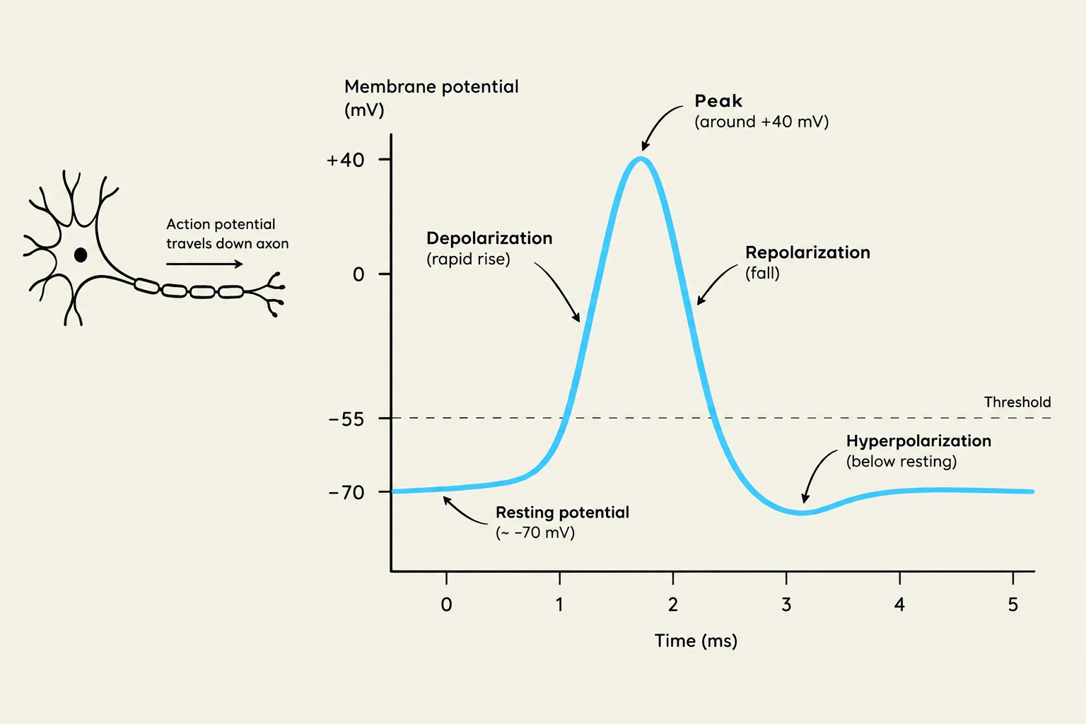
The cycle begins when a stimulus causes the membrane potential to reach a specific "threshold." Once this threshold is met, voltage-gated sodium (Na+) channels open rapidly, allowing an influx of positive ions into the cell. This process, known as depolarization, makes the inside of the neuron much more positive than the outside.
To prevent the cell from remaining in this excited state, the neuron must reset. This is achieved through repolarization, where sodium channels close and voltage-gated potassium ($K^+$) channels open. Potassium ions flow out of the cell (efflux), removing positive charge and returning the membrane potential toward its resting state. Finally, the Na+/K+ ATPase pump works to restore the original ionic concentrations, ensuring the neuron is ready for the next signal.
A graph illustrating the changes in membrane potential during an action potential in a neuron, showing resting potential, depolarization, peak, repolarization, and hyperpolarization phases.
The All-or-None Principle and Refractory Periods
A fundamental rule of neural signaling is the all-or-none principle: an action potential does not vary in strength. If the threshold is reached, a full-amplitude impulse is generated; if the threshold is not reached, nothing happens.
To ensure that signals travel in only one direction and to prevent the neuron from being overwhelmed, the system utilizes refractory periods:
Absolute Refractory Period: A time during which it is physically impossible for the neuron to fire another action potential, regardless of the stimulus strength.
Relative Refractory Period: A period following the absolute phase where a second action potential is possible, but only if the stimulus is significantly stronger than usual.
Speeding Up the Signal: Saltatory Conduction
Not all signals travel at the same speed. The velocity of an impulse depends on axon diameter and the presence of myelin (a fatty insulating layer). In myelinated axons, the electrical impulse "jumps" from one gap in the myelin (Node of Ranvier) to the next. This process is called saltatory conduction, and it allows for much faster communication than continuous conduction in unmyelinated fibers.
Exam Tip: If a question asks why a reflex is faster than a slow sensory modulation, look for "myelination" and "saltatory conduction" in your reasoning.
Chemical Transmission: The Synapse
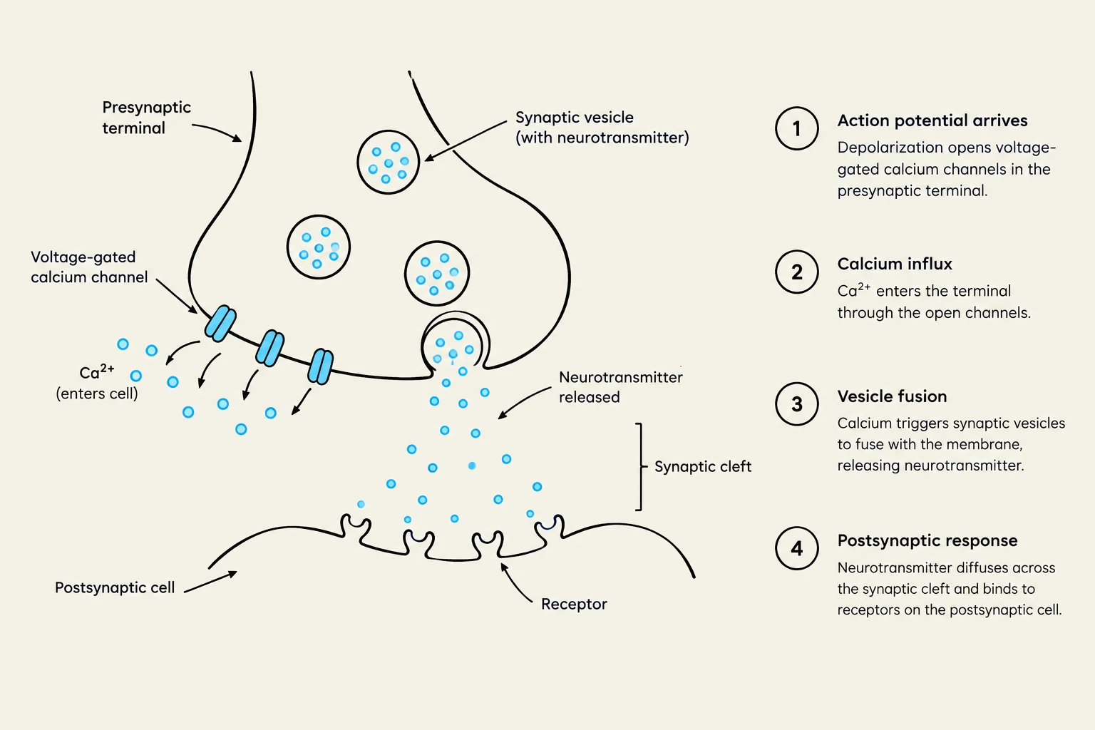
When the electrical action potential reaches the end of an axon (the terminal button), it must cross a gap called the synaptic cleft to reach the next cell. This is where electricity is converted into chemistry.
Neurotransmitter Release and Receptors
The arrival of the action potential triggers an influx of calcium ions (Ca2+) into the terminal. This calcium influx acts as a signal for vesicles to fuse with the membrane and release neurotransmitters into the cleft. These chemicals then bind to receptors on the postsynaptic neuron, which fall into two main categories:
Ionotropic Receptors: These are ligand-gated ion channels. When the neurotransmitter binds, the channel opens immediately, allowing direct ion flow. This results in very fast signaling.
Metabotropic Receptors: These are slower and more complex. They do not form a channel themselves but instead activate "second messenger" pathways inside the cell. While slower, they can produce much more widespread and long-lasting effects.
Diagram illustrating the process of synaptic transmission, including action potential arrival, calcium influx, vesicle fusion, and postsynaptic response.
To prevent constant stimulation, the signal is terminated through three methods: reuptake by the sending neuron, enzymatic breakdown of the chemical, or simple diffusion away from the site.
Integration and Plasticity
A single neuron is often receiving thousands of signals simultaneously. It must "decide" whether to fire its own action potential based on the sum of these inputs.
EPSPs, IPSPs, and Summation
Incoming signals are categorized by their effect on the postsynaptic membrane:
EPSPs (Excitatory Postsynaptic Potentials): These depolarize the membrane, bringing the neuron closer to the threshold.
IPSPs (Inhibitory Postsynaptic Potentials): These hyperpolarize the membrane, making it more negative and moving it further from the threshold.
The neuron performs integration through two types of summation:
Temporal Summation: Multiple signals arriving at the same synapse in rapid succession.
Spatial Summation: Signals arriving from many different synapses across the cell body at the same time.
Synaptic Plasticity: The Basis of Learning
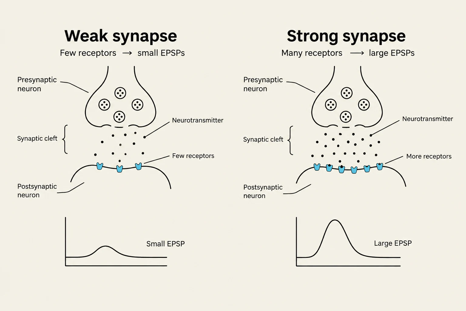
The nervous system is not hard-wired. Through synaptic plasticity, the strength of the connections between neurons can change based on activity. If two neurons fire together repeatedly, their connection may strengthen (making EPSPs more effective); if they fire out of sync, the connection may weaken. This ability to modify connections is the biological foundation of learning, memory, and adaptation.
Diagram illustrating the difference between a weak synapse with few receptors and a strong synapse with many receptors, affecting the size of excitatory postsynaptic potentials (EPSPs).
Summary Table: Quick Review
Feature
Function
Action Potential
Rapid electrical signal along an axon
Neurotransmitter
Chemical messenger across a synapse
EPSP
Excitatory; moves membrane toward threshold
IPSP
Inhibitory; moves membrane away from threshold
Plasticity
Change in synaptic strength; enables learning
Keep reading for free.
Create an account to finish this reading and make it part of your study routine.