Neurological Exam and Diagnostic Tests: A Guide for Healthcare Professionals
This guide explores the multifaceted approach to neurological assessment, from initial bedside exams to advanced imaging and electrophysiological testing, emphasizing the interdisciplinary team's role in diagnosing neurological conditions.
When a patient presents with neurological symptoms—such as sudden weakness, confusion, or loss of sensation—the medical team must act quickly to identify the underlying cause. The process of determining what is wrong involves a combination of clinical observation and advanced technology. This article explores the multifaceted approach to neurological assessment, ranging from the initial bedside exam to complex imaging and electrophysiological testing, highlighting how different healthcare professionals contribute to the diagnostic puzzle.
The Neurological Exam: From Screening to In-Depth Assessment
The neurological exam is not a single event but a series of tests and observations designed to evaluate the functional status of the nervous system. It serves as a critical piece of clinical decision-making, often used in conjunction with medical diagnostic tests to confirm a suspected diagnosis or rule out other possibilities.
The Rapid Neurological Screen
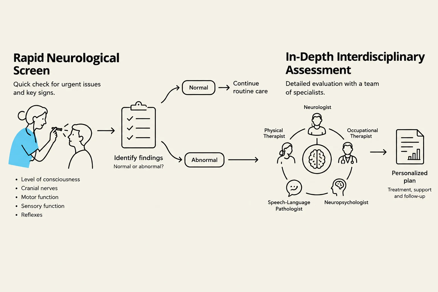
Typically, a neurologist begins with a rapid screening to check major neural functions. This high-level overview includes:
Mental Status: Assessing alertness, language, attention, recent memory, and orientation to time and place.
Cranial Nerves: Checking pupil symmetry, eye movements, and facial, neck, or tongue movements.
Reflexes: Testing upper and lower limbs for symmetry and checking for the return of primitive reflexes (e.g., rooting).
Motor System: Observing movement, muscle tone, and strength.
Coordination and Gait: Using tests like "finger-to-nose" to check for tremors and observing walking ability or transfers.
Sensory System: Using verbal reports (pain/numbness) and physical tests (cotton, pin, or vibration).
This diagram illustrates the process of a rapid neurological screen and an in-depth interdisciplinary assessment, leading to a personalized treatment plan.
The Role of OT and PT in In-Depth Examination
If the initial screen reveals abnormalities, specialized professionals like Occupational Therapists (OT) and Physical Therapists (PT) perform a much more granular, functional assessment. While a neurologist focuses on the physiological "hardware," OTs and PTs focus on how neurological deficits impact daily life and movement.
Cognitive and Perceptual Function: OTs assess how cognition impacts function, looking at learning, memory, attention, and "neglect" (ignoring one side of space). They may use tools like the MoCA or evaluate complex tasks like planning and deciding.
Motor and Sensory Function: PTs and OTs dive deep into the motor system. This includes inspecting for muscle atrophy, testing range of motion (goniometry), and assessing muscle tone using scales like the Modified Ashworth Scale to identify hypertonia or spasticity. They also evaluate coordination through specific tests like the heel-shin or foot tap tests, and assess sensory nuances such as proprioception (body position awareness) and stereognosis (identifying objects by touch).
Clinical Tip: Always view the neurological exam within the context of the patient's history and initial interview. The exam provides the "what," but the history provides the "why".
Brain and Spinal Cord Imaging
When clinical exams suggest structural issues, medical teams turn to imaging to visualize the brain and spinal cord.
Computed Tomography (CT Scan)
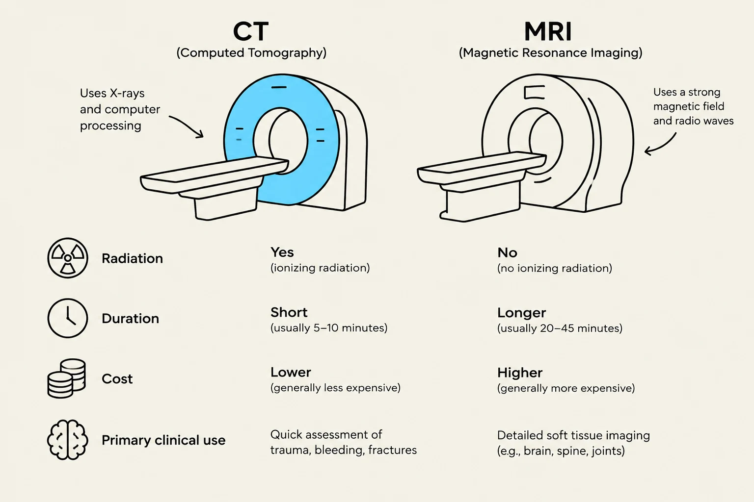
A CT scan is a non-invasive imaging method that uses computer processing technology and limited radiation. It is relatively fast, typically lasting only 5–10 minutes.
Capabilities: It can differentiate grey matter from white matter and visualize the ventricles.
Primary Uses: CT is essential for detecting bleeding (hemorrhage), brain damage, blood clots, or leaking aneurysms. It is also used to detect most brain tumors, identify enlarged ventricles in hydrocephalus, and guide needles during a biopsy.
Magnetic Resonance Imaging (MRI)
MRI provides much higher resolution than a CT scan by using magnets to influence magnetic fields along three spatial axes.
Capabilities: It creates highly detailed spatial images without using radiation.
Limitations: It is more costly, takes longer (30–45 minutes), and cannot be used if the patient has metal-containing implants, such as a pacemaker.
Primary Uses: MRI is the gold standard for localizing strokes, tumors, and traumatic injuries with significant detail. It is also used to assess diffuse brain changes or atrophy associated with dementia, Parkinson’s, or multiple sclerosis.
Comparison of CT and MRI imaging techniques, highlighting differences in radiation use, duration, cost, and primary clinical applications.
Advanced Imaging: PET and fMRI
Beyond standard structural imaging, specialized scans provide functional data:
Positron Emission Tomography (PET): Used to observe metabolic processes in the brain.
Functional Magnetic Resonance Imaging (fMRI): Measures brain activity by detecting changes associated with blood flow.
Electrophysiological and Other Diagnostic Tests
To understand the electrical and chemical health of the nervous system, clinicians use several other specialized tests.
Electrophysiological Measurements
These tests measure the "signals" traveling through the nervous system:
Electroencephalography (EEG): Uses electrodes on the scalp to measure cortical activity. It is frequently used to study epilepsy and sleep disorders. Interestingly, research has shown that EEG indices can even help predict a patient's recovery of consciousness from a coma.
Evoked Potentials (EP): These measure the electrical response of the nervous system to specific stimuli to test the integrity of sensory or motor pathways.
Electromyography (EMG): This tests the electrical activity within a muscle to evaluate the strength and speed of the signal coming from the nerve.
Invasive and Laboratory Tests
Lumbar Puncture (CSF Test): A procedure to collect cerebrospinal fluid for analysis, which can reveal infections, inflammation, or other abnormalities.
Biopsy: A tissue sample may be taken to provide a definitive diagnosis of a tumor or other pathology. CT scans are often used to guide the needle during this process.
Blood and Urine Tests: Used to rule out metabolic or systemic causes of neurological symptoms.
Summary for the Interdisciplinary Team
For students and practitioners in Physical and Occupational Therapy, understanding these tests is vital. The neurological exam is not just the doctor's responsibility; it is a collaborative process. Abnormalities found during a PT or OT session are brought back to the interdisciplinary team, which may then order further medical diagnostic tests to refine the diagnosis and treatment plan.
By integrating the functional observations of therapy with the structural and electrical data from medical imaging and electrophysiology, the team can provide comprehensive care for patients with neurological conditions.
Keep reading for free.
Create an account to finish this reading and make it part of your study routine.