A systematic evaluation of the nervous system, including level of consciousness, language, cranial nerve status, movement, sensation, and vital signs. Covers the twelve cranial nerves, their origins, and functions, as well as clinical correlations with conditions like stroke, Parkinson's disease, and Multiple Sclerosis
A neurologic assessment is a systematic evaluation used to determine the status of a patient's nervous system. For an awake and alert patient, this process involves a comprehensive review of their level of consciousness (LOC), language and speech, cranial nerve status, movement and sensation, and vital signs. In cases where a patient exhibits altered consciousness, clinicians must rely on more structured tools like the Glasgow Coma Scale (GCS) to quantify impairment and monitor motor responses.
The Fundamentals of Neurologic Assessment
When assessing a patient, the clinician must look beyond immediate physical signs and gather a complete history. This includes understanding any physical disabilities, nutritional status, and family history or genetic risks.
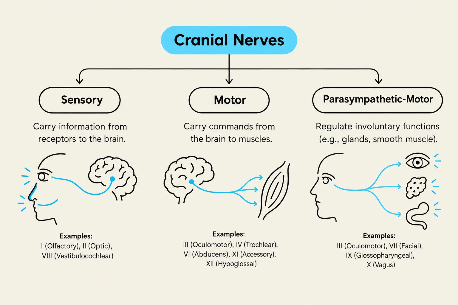
The assessment is often categorized by the type of neurological function being tested:
Sensory Perception: The ability to process stimuli such as touch, temperature, pain, and specialized senses like sight and smell.
Motor Functions: The ability to execute voluntary and involuntary movements, including muscle strength and coordination.
Parasympathetic-Motor: The involuntary control of smooth muscles and glands, such as pupil constriction or salivary secretion.
Exam Tip: When assessing a patient with altered consciousness, the Glasgow Coma Scale (GCS) is the gold standard for quantifying neurological impairment. Always prioritize the assessment of the airway and vital signs alongside the GCS score.
The Twelve Cranial Nerves
The cranial nerves are a set of twelve pairs of nerves that emerge directly from the brain and brainstem. They are essential for sensory perception and motor control of the head, neck, and visceral organs. Understanding their origin and specific function is critical for localizing neurological lesions.
Sensory and Motor Mapping
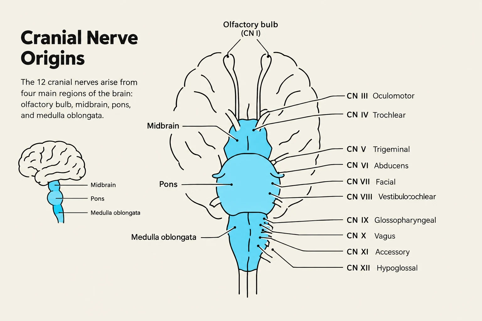
The nerves originate from different levels of the brainstem, including the olfactory bulb, the midbrain, the pons, and the medulla.
Nerve
Origin
Type
Primary Function
I: Olfactory
Olfactory bulb
Sensory
Smell
II: Optic
Midbrain
Sensory
Central and peripheral vision
III: Oculomotor
Midbrain
Motor/Parasympathetic
Eye movement, lid elevation, pupil constriction
IV: Trochlear
Lower midbrain
Motor
Eye movement (superior oblique)
V: Trigeminal
Pons
Sensory/Motor
Facial sensation and chewing (mastication)
VI: Abducens
Inferior pons
Motor
Eye movement (lateral rectus)
VII: Facial
Inferior pons
Sensory/Motor/Para
Facial expression, taste (ant. 2/3), glands
VIII: Vestibulocochlear
Pons-medulla junction
Sensory
Hearing and equilibrium
IX: Glossopharyngeal
Medulla
Sensory/Motor/Para
Taste/sensation (post. 1/3), throat muscles
X: Vagus
Medulla
Sensory/Motor/Para
Swallowing, sensation in viscera, parasympathetic
XI: Accessory
Medulla/Cervical spine
Motor
Sternocleidomastoid and trapezius muscles
XII: Hypoglossal
Medulla
Motor
Tongue movement for swallowing
Diagram illustrating the origins of the 12 cranial nerves from different regions of the brain, including the olfactory bulb, midbrain, pons, and medulla oblongata.
Detailed Nerve Functions
The Midbrain and Pons Connection
The midbrain is a critical center for visual and ocular motor control, housing the origins for the Optic (II), Oculomotor (III), and Trochlear (IV) nerves. Moving down to the pons, we find the Trigeminal (V) and Facial (VII) nerves, which manage everything from facial sensation to the muscles used for chewing and expression. The Abducens (VI) nerve also originates in the inferior pons to assist with lateral eye movement.
The Medulla and Vital Functions
The medulla serves as the origin for several high-stakes nerves. The Glossopharyngeal (IX), Vagus (X), Accessory (XI), and Hypoglossal (XII) nerves all emerge here. The Vagus nerve is particularly notable for its extensive parasympathetic-motor role, regulating sensation in the pharynx and larynx, controlling swallowing, and providing innervation to the thoracic and abdominal viscera.
This diagram illustrates the three types of cranial nerves: sensory, motor, and parasympathetic-motor, along with their functions and examples.
Clinical Correlations: When Assessment Reveals Pathology
A deficit in a specific cranial nerve or a change in the level of consciousness can point toward serious underlying conditions such as stroke, Parkinson's disease, or Multiple Sclerosis.
Stroke and Increased Intracranial Pressure (ICP)
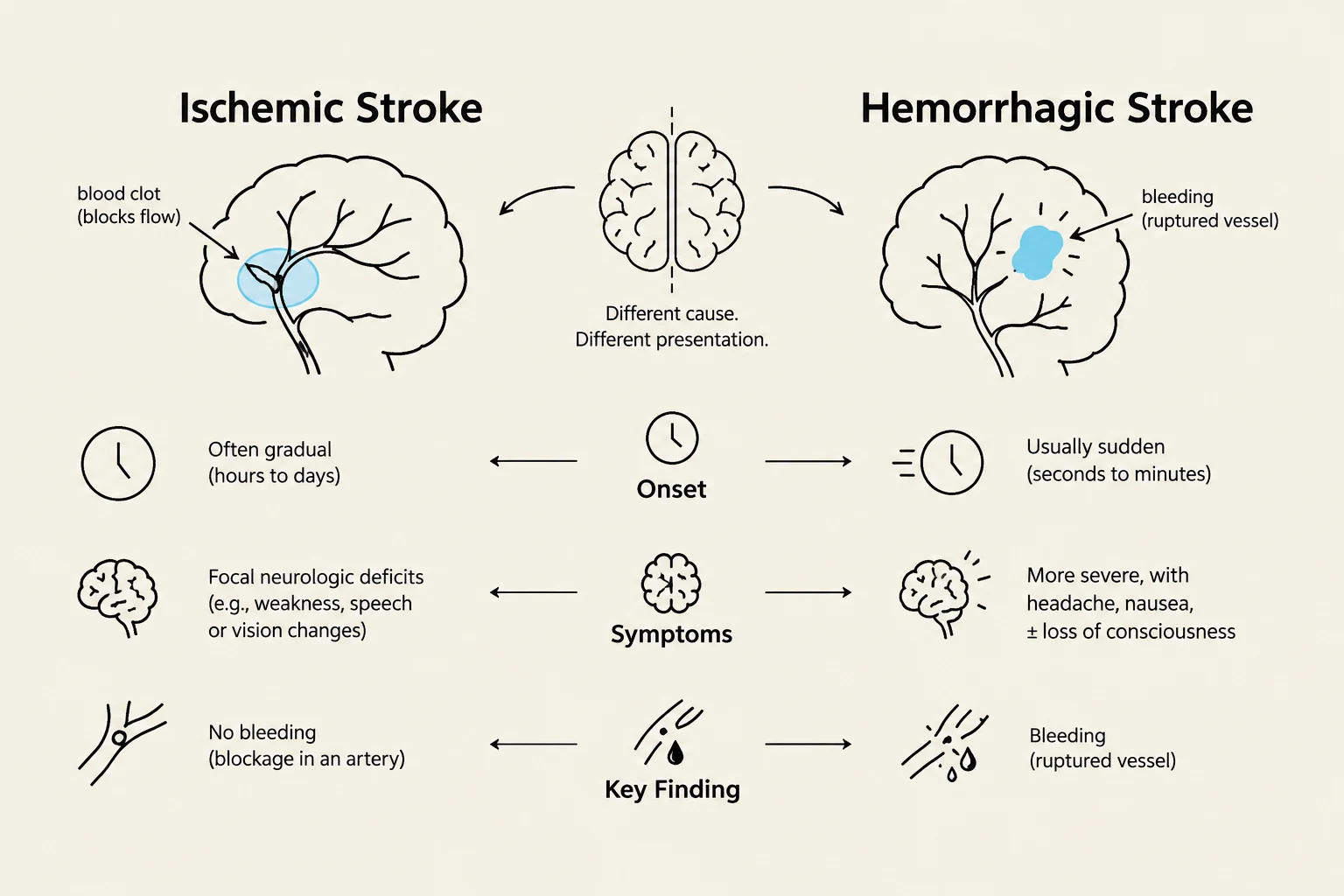
A stroke is an interruption of blood perfusion to the brain, resulting in cell death (infarction). This can be Ischemic (blockage) or Hemorrhagic (bleeding).
One of the most dangerous complications of a stroke is Increased Intracranial Pressure (ICP). Clinicians must watch for the "Cushing triad," a very late sign of increased pressure consisting of:
Severe hypertension
Widened pulse pressure
Bradycardia
Clinical Alert: The earliest sign of increased ICP is often a decreased level of consciousness (LOC). If a patient shows sudden behavioral changes, headache, or projectile vomiting, immediate intervention is required.
Parkinson's Disease and Motor Deficits
In Parkinson's disease, the degradation of dopamine in the substantia nigra leads to profound motor dysfunction. Key "cardinal signs" to look for during a motor assessment include:
Bradykinesia: Slowness of movement.
Resting Tremor: Often described as "pill rolling".
Rigidity: Stiffness of the extremities.
Postural Instability: Difficulty with balance and gait.
Multiple Sclerosis (MS) and Sensory/Motor Disruption
Multiple Sclerosis is an autoimmune, inflammatory disease that affects the myelin sheath. Because demyelination can occur anywhere along the brain, optic nerve, or spinal cord, patients present with a wide array of sensory and motor symptoms. Common findings include:
Sensory: Tinnitus, blurred vision, diplopia (double vision), and paresthesia (numbness/tingling).
Motor: Muscle spasticity, tremors, ataxia (lack of coordination), and unsteady gait.
This diagram illustrates the differences between ischemic and hemorrhagic strokes, detailing their causes, onset, symptoms, and key findings.
Summary of Assessment Priorities
When performing a neurologic assessment, remember that the goal is to localize the problem. Is it a sensory issue (e.g., Olfactory or Optic)? A motor issue (e.g., Trigeminal or Hypoglossal)? Or a global issue involving the level of consciousness (e.g., GCS)? By systematically evaluating these components, clinicians can provide life-saving interventions for acute events like strokes or seizures.
Keep reading for free.
Create an account to finish this reading and make it part of your study routine.