Neuroimaging Techniques: Visualizing Brain Structure and Function
An overview of neuroimaging techniques, including structural methods like CT, MRI, and DTI, and functional methods such as PET and fMRI. Learn how these tools help scientists study the brain's anatomy and activity.
For much of human history, the brain was a "black box"—an organ whose internal workings could only be inferred from behavior or observed after death through autopsy. Today, neuroimaging has revolutionized neuroscience by allowing us to peer inside the living brain in real-time. By using various physical principles—ranging from X-rays to magnetic fields and radioactive tracers—scientists can distinguish between the physical anatomy of the brain and the dynamic processes of thought, emotion, and perception.
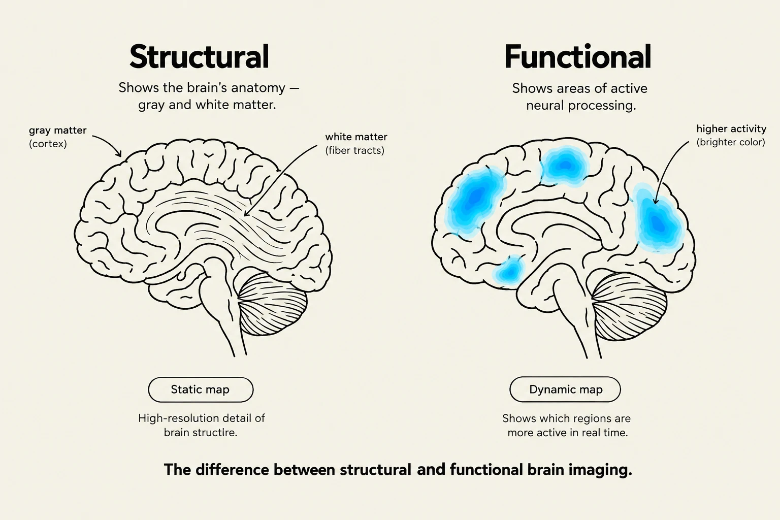
The difference between structural and functional brain imaging, illustrating brain anatomy and areas of neural processing.
Understanding these techniques requires a fundamental distinction between structural imaging (what the brain looks like) and functional imaging (what the brain is doing).
Structural Imaging: Mapping the Anatomy
Structural imaging techniques are used to visualize the physical shape, size, and integrity of brain tissues. These are essential for identifying tumors, hemorrhages, or physical damage caused by trauma.
Computed Tomography (CT)
The CT scan is one of the most common medical imaging tools. It uses a series of X-ray measurements taken from different angles around the head to create cross-sectional images (slices) of the brain.
Best Use Case: Emergency rooms. Because CT scans are fast and highly effective at detecting bone fractures and acute bleeding (hemorrhages), they are often the first line of defense in trauma cases.
Limitation: It involves exposure to ionizing radiation, which can be a concern with repeated use.
Magnetic Resonance Imaging (MRI)
MRI provides much higher resolution than a CT scan. Instead of X-rays, it uses powerful magnetic fields and radio waves to manipulate the protons in the body's water molecules. When the magnetic field is turned off, these protons release energy that is captured to create highly detailed images.
Best Use Case: Detailed anatomical studies. If a researcher needs to see the exact boundary between gray matter and white matter, or if a doctor needs to look for small lesions, MRI is the gold standard.
Limitation: It is much slower than a CT scan and requires the patient to remain perfectly still inside a loud, enclosed tube.
Diffusion Tensor Imaging (DTI)
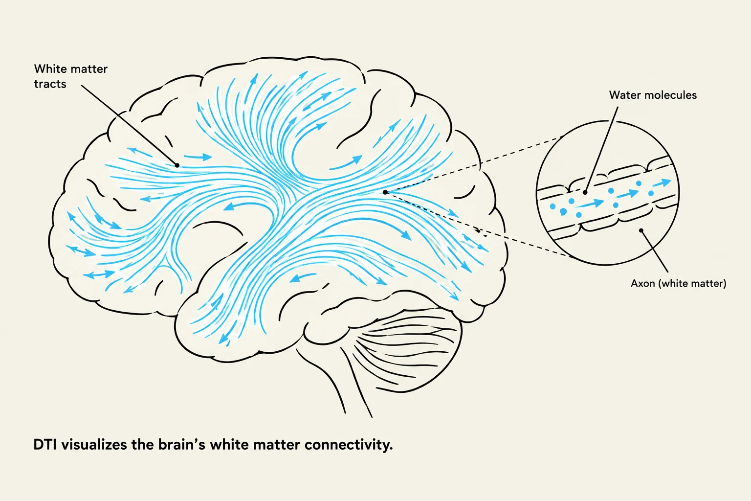
This diagram illustrates the brain's white matter connectivity using Diffusion Tensor Imaging (DTI) to visualize water molecule movement along axons.
DTI is a specialized type of MRI that goes a step further by mapping the movement of water molecules within the brain's white matter. Because water moves more easily along the length of axons than across them, DTI can visualize the "wiring" or connectivity of the brain.
Best Use Case: Studying "connectomics." DTI helps researchers understand how different brain regions communicate via white matter tracts.
Exam Tip: If a question asks about the difference between CT and MRI, remember: CT = Speed/Emergency/X-rays; MRI = Detail/Anatomy/Magnets.
Functional Imaging: Mapping Brain Activity
While structural imaging shows us the "hardware," functional imaging shows us the "software" in action. These techniques track the physiological changes that occur when neurons fire.
Positron Emission Tomography (PET)
PET scans involve injecting a small amount of a radioactive tracer (often a form of glucose) into the bloodstream. Since active neurons consume more energy, they will take up more of this tracer. The PET scanner detects the radiation emitted by the tracer to create a map of metabolic activity.
Best Use Case: Studying neurochemistry and disease progression. PET can be used to see how certain drugs interact with receptors or how glucose metabolism changes in Alzheimer's disease.
Limitation: It is invasive due to the radioactive tracer and has relatively low temporal resolution (it shows what happened over several minutes, not milliseconds).
Functional Magnetic Resonance Imaging (fMRI)
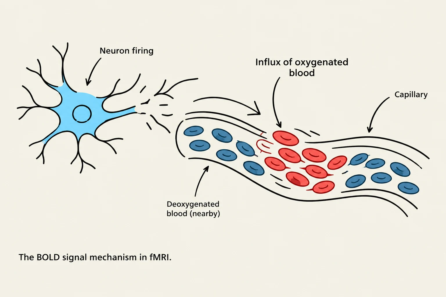
The BOLD signal mechanism in fMRI illustrates how neural activity leads to an influx of oxygenated blood into capillaries, changing the magnetic properties detected by fMRI.
fMRI is currently the most popular tool in cognitive neuroscience. It relies on the BOLD (Blood-Oxygen-Level Dependent) signal. When a specific brain region becomes active, it requires more oxygen. The body responds by sending a surge of oxygenated blood to that area. Because oxygenated and deoxygenated blood have different magnetic properties, the MRI machine can detect these changes.
Best Use Case: Mapping cognitive functions. For example, a researcher can ask a participant to look at faces while in the scanner to see which specific areas of the brain "light up" during facial recognition.
Limitation: It measures blood flow, which is an indirect proxy for neural activity. There is a slight delay between a neuron firing and the blood flow responding.
Summary Comparison Table
To help synthesize these concepts, the following table compares the primary characteristics of each technique:
Technique
Primary Use
Mechanism
Main Advantage
CT
Structural
X-rays
Fast; great for emergencies
MRI
Structural
Magnetic fields
High anatomical detail
DTI
Structural
Water diffusion
Visualizes white matter tracts
PET
Functional
Radioactive tracers
Measures metabolic/chemical activity
fMRI
Functional
Blood oxygen levels
Non-invasive; maps brain activity
Conclusion
Choosing the right neuroimaging tool depends entirely on the research question or medical need. If the goal is to check for a brain bleed after a car accident, a CT is the fastest and most effective choice. If a scientist wants to know which part of the brain processes language, fMRI provides the necessary functional map. By combining these technologies, modern neuroscience continues to peel back the layers of the human mind, moving from the physical structure of the brain to the complex functions that define our existence.
Keep reading for free.
Create an account to finish this reading and make it part of your study routine.