


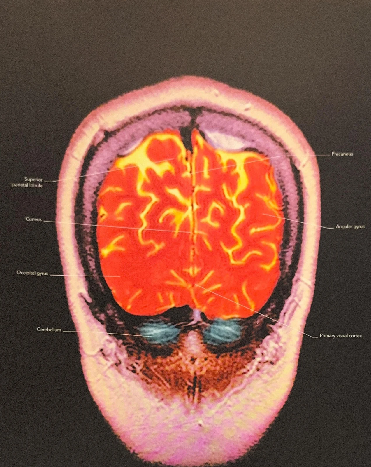
Understanding the human brain requires a journey through various specialized structures, each serving as a critical node in a vast, interconnected network. While some regions are famous for their obvious roles in movement or sight, others remain shrouded in mystery, playing subtle but vital roles in how we perceive ourselves and our history. This article explores the neuroanatomy and functional specializations of the cerebellum, the occipital lobe, the primary visual cortex, and the midline structures of the precuneus and posterior cingulate cortex.
The Cerebellum: The Architect of Movement
Located at the back and below the main cerebrum, the cerebellum is often referred to as the "little brain" due to its distinct structure and position. Despite its smaller size relative to the cerebrum, it is highly convoluted, containing a massive density of neurons that allow for complex processing.
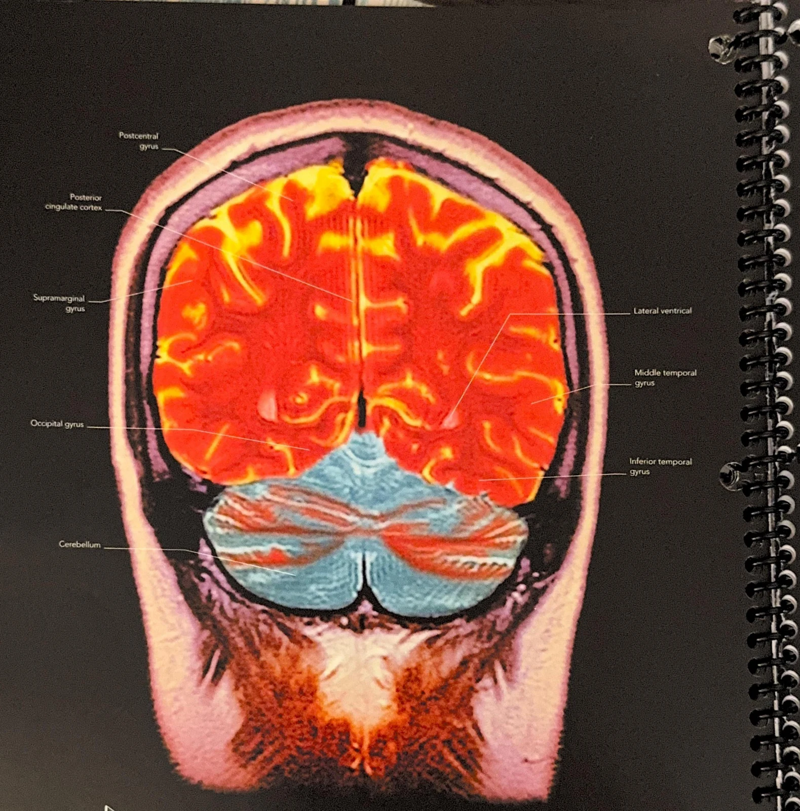
The primary functional domain of the cerebellum is fine motor control and the timing of movements. While the motor cortex initiates movement, the cerebellum acts as a real-time coordinator. It maintains extensive connections with the motor cortex to ensure that physical activities are smooth, accurate, and well-timed.
Student Tip: Coordination vs. Initiation A common mistake is to confuse the roles of the motor cortex and the cerebellum. Remember: the motor cortex is responsible for the command to move, whereas the cerebellum is responsible for the refinement and precision of that movement. Damage to the cerebellum typically results in "ataxia"—clumsy, uncoordinated movements—rather than total paralysis.


Keep reading for free.
Create an account to finish this reading and make it part of your study routine.
Continue reading freeAlready have an account? Log in
Updated