An overview of the nervous system's structure, function, and common diseases. Covers the Central and Peripheral Nervous Systems, brain anatomy, neurophysiology, and conditions like Multiple Sclerosis and Parkinson's Disease.
The nervous system is a sophisticated, continuous loop of sensory input, integration, and motor output. It allows an organism to perceive its environment, process that information, and execute appropriate physical responses. To understand how this system functions—and why it fails in diseases like Multiple Sclerosis or Parkinson’s—one must understand the interplay between its structural divisions and its chemical communication methods.
Structural Organization: CNS and PNS
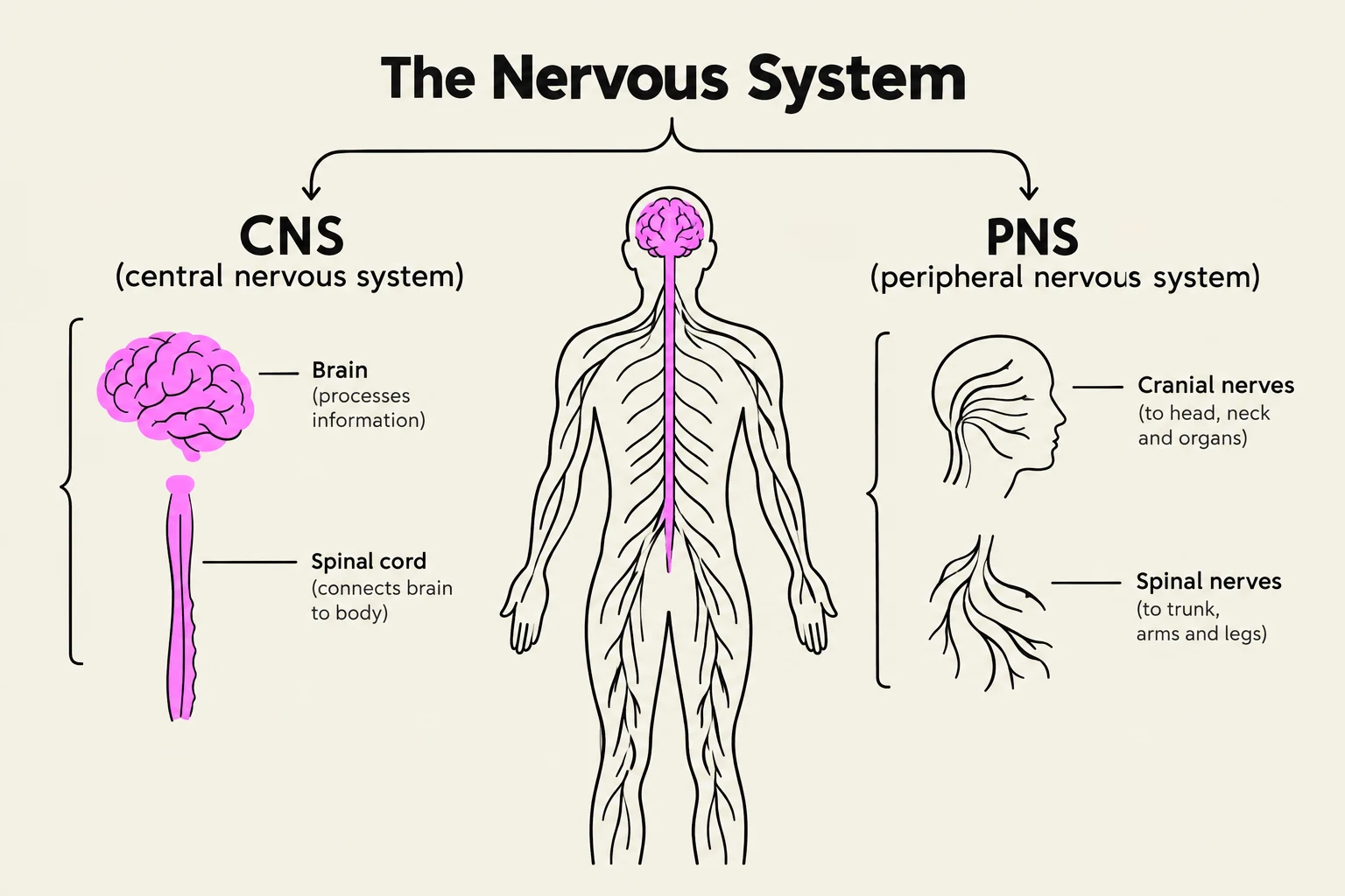
The nervous system is divided into two primary anatomical domains: the Central Nervous System (CNS) and the Peripheral Nervous System (PNS).
The Central Nervous System (CNS)
The CNS serves as the integration center, consisting of the brain and the spinal cord. The brain is a complex structure containing the cerebrum, diencephalon, brainstem, and cerebellum. The spinal cord acts as a high-speed highway for ascending sensory tracts and descending motor tracts, while also serving as the center for reflex arcs. Because the CNS is protected by the skull, vertebrae, meninges, and the blood-brain barrier, it has a very limited capacity to regenerate following injury.
The Peripheral Nervous System (PNS)
The PNS consists of the nerves that connect the CNS to the rest of the body. This includes 12 pairs of cranial nerves that arise directly from the brain and brainstem, and 31 pairs of spinal nerves. Unlike the CNS, the PNS possesses a greater capacity for regeneration, largely due to the presence of Schwann cells which produce myelin.
The PNS is further divided into two functional systems:
Somatic Division: Manages voluntary control of skeletal muscles and provides sensory input.
Autonomic Division: Manages involuntary functions, such as the heart rate and glandular secretions.
Clinical Note: Dermatomes
Each spinal nerve serves a specific area of skin known as a dermatome. In clinical practice, if a patient reports numbness in a specific pattern, a clinician can map that sensation back to a specific spinal nerve level to locate a lesion.
Diagram illustrating the central nervous system (CNS) and peripheral nervous system (PNS), detailing the brain, spinal cord, cranial nerves, and spinal nerves.
Functional Anatomy of the Brain
The brain is not a uniform mass; different regions are specialized for specific tasks:
Frontal Lobe: Responsible for voluntary movement, judgment, personality, and expressive speech (the Broca area).
Parietal Lobe: Handles sensory integration, touch, and spatial awareness.
Temporal Lobe: Manages hearing, memory, and language comprehension (the Wernicke area).
Occipital Lobe: Dedicated to visual processing and interpretation.
Cerebellum: Essential for coordination, balance, and the fine-tuning of movement. Damage here can lead to ataxia and intention tremors.
Brainstem: Includes the midbrain, pons, and medulla; it controls vital life functions like heart rate and breathing.
Basal Ganglia: Deep brain nuclei that initiate and smooth out voluntary movements.
Neurophysiology: Signals and Communication
Effective nervous system function relies on the movement of electrical impulses and chemical messengers.
Electrical Conduction and Myelin
Signals travel along axons as an "all-or-none" electrical impulse called an action potential. This involves depolarization (sodium ions Na+ rushing into the cell) and repolarization (potassium ions $K^+$ exiting the cell).
To speed up this process, many axons are wrapped in myelin, a lipid-based insulation. In myelinated axons, the impulse does not move like a continuous wave; instead, it "jumps" from one Node of Ranvier to the next, a process called saltatory conduction. If myelin is lost—a process known as demyelination—the signal may slow down or fail entirely.
Chemical Transmission: Synapses and Neurotransmitters
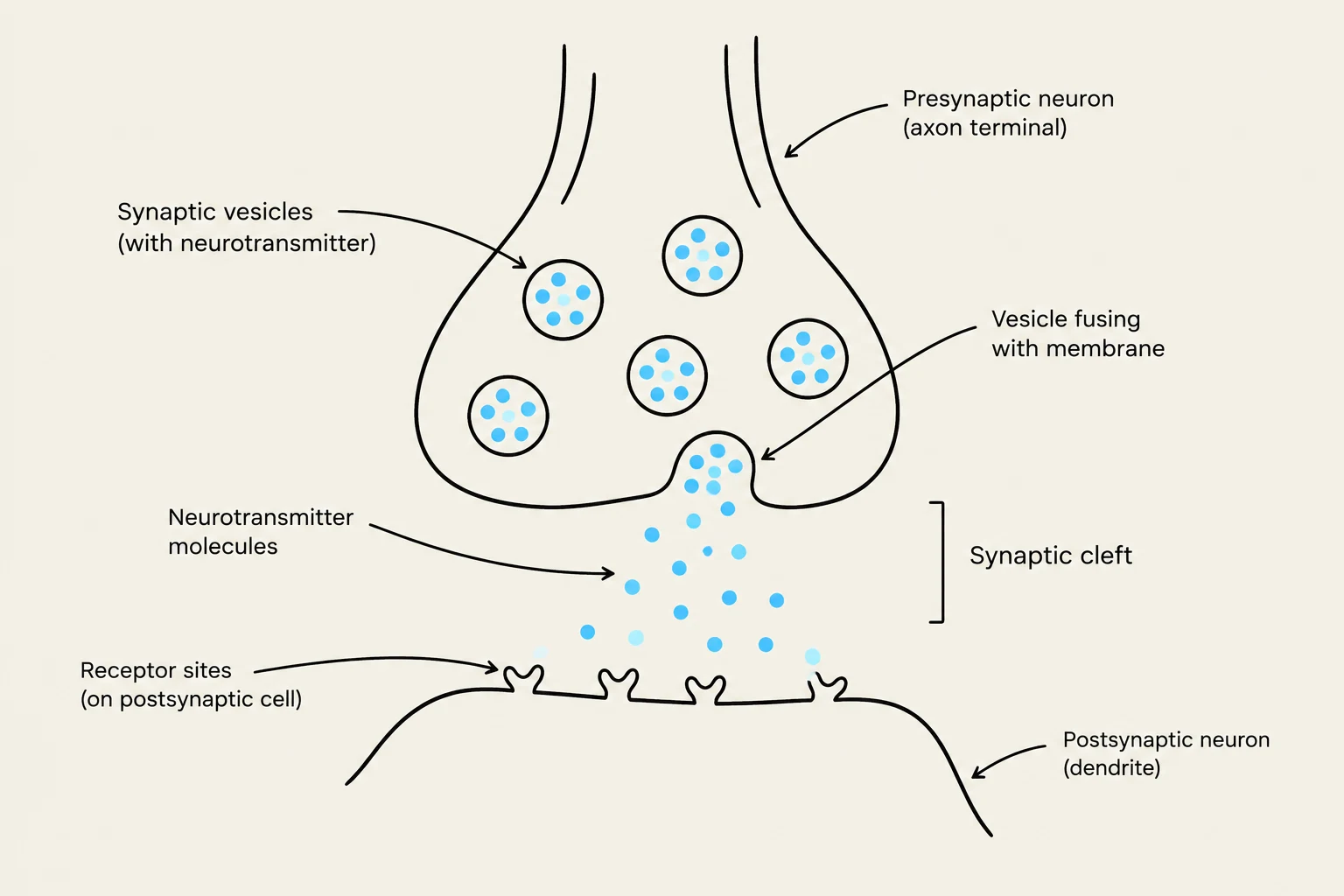
When an electrical impulse reaches the end of an axon (the terminal), it must cross a gap to reach the next cell. This junction is the synapse. To bridge this gap, the neuron releases neurotransmitters, which are chemical messengers that bind to specific receptors on the receiving (post-synaptic) cell.
Diagram illustrating a synapse, the junction between two neurons where information is transmitted.
The Autonomic Nervous System (ANS)
The ANS regulates involuntary functions through two opposing systems that work together to maintain homeostasis:
Sympathetic Nervous System ("Fight or Flight"): Prepares the body for stress or intense activity. It originates in the thoracolumbar region (T1 to L2) and primarily uses norepinephrine as its transmitter.
Parasympathetic Nervous System ("Rest and Digest"): Promotes energy conservation and maintenance functions. It originates in the craniosacral region (Cranial nerves III, VII, IX, X and sacral S2 to S4) and primarily uses acetylcholine (ACh).
The Two-Neuron Chain
The autonomic pathway utilizes a unique two-neuron chain:
Preganglionic neuron: Located within the CNS.
Synapse: Occurs within a peripheral ganglion.
Postganglionic neuron: Extends from the ganglion to the target organ.
Exam Tip: Acetylcholine (ACh)
Remember that ACh is released at all autonomic ganglia. However, the neurotransmitter released at the target organ changes depending on whether the system is sympathetic or parasympathetic.
Pathophysiology: When Systems Fail
Neurological diseases can be categorized by whether they represent a failure of "insulation" or a failure of "chemical messengers".
Multiple Sclerosis (MS): Insulation Failure
MS is an immune-mediated disease where the body's immune system attacks the myelin sheath in the CNS. This results in the formation of "plaques" (scarred, demyelinated lesions) in the white matter.
Clinical Manifestations: Symptoms vary by location but often include fatigue (the most common), optic neuritis (visual issues), ataxia (cerebellar issues), and Lhermitte sign (an electric shock sensation down the spine).
Uhthoff Phenomenon: A unique occurrence where MS symptoms temporarily worsen when core body temperature rises (e.g., during exercise or fever) due to slowed conduction in compromised axons.
Parkinson Disease (PD): Chemical Failure
While MS is an insulation problem, Parkinson Disease is a problem with a chemical messenger—specifically dopamine. PD involves the progressive death of dopaminergic neurons in the substantia nigra. This creates an imbalance where there is a relative excess of Acetylcholine.
The TRAP Acronym: Motor symptoms are often summarized as:
Tremor (pill-rolling resting tremor)
Rigidity ("cogwheel" muscle stiffness)
Akinesia/Bradykinesia (slowness or difficulty initiating movement)
Postural Instability (impaired balance and shuffling gait)
Peripheral Neuropathies and Neuromuscular Disease
Pathology can also occur in the peripheral nerves or the junction where nerves meet muscles:
Guillain-Barré Syndrome (GBS): An acute, autoimmune demyelinating polyneuropathy of the PNS that often presents as ascending, symmetrical muscle weakness.
Myasthenia Gravis (MG): An autoimmune disorder of the neuromuscular junction where antibodies block or destroy Acetylcholine receptors (AChR) on the muscle cell. This leads to muscle weakness that worsens with activity and improves with rest.
Keep reading for free.
Create an account to finish this reading and make it part of your study routine.