An explanation of the nervous system's fundamental electrical processes, including resting membrane potential, graded potentials, action potentials, and synaptic transmission.
The nervous system is a complex communication network designed to sense environmental changes, process information, and trigger appropriate reactions. At the heart of this system is the neuron, the functional unit responsible for transmitting electrical impulses. To understand how a thought is formed or a muscle is moved, we must look at the microscopic electrical events occurring across the neuronal membrane.
The Resting Membrane Potential
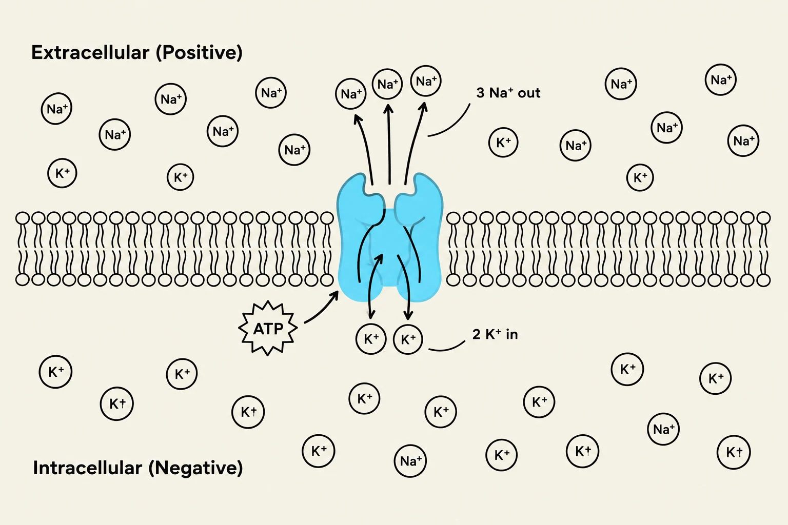
Diagram illustrating the sodium-potassium pump, a transmembrane protein that moves sodium ions out of the cell and potassium ions into the cell, utilizing ATP for energy.
Before a neuron can send a signal, it must be in a state of readiness. This state is known as the resting membrane potential (RMP). The RMP represents a relative difference in electrical charges across the plasma membrane.
In a resting neuron, the extracellular region (outside the cell) is more positive, while the intracellular region (inside the cell) is more negative. This electrical gradient is maintained by several key mechanisms:
Ion Leak Channels: These allow ions to move slowly across the membrane.
Sodium–Potassium Exchange Pump: This is an active process that uses ATP to move ions against their concentration gradients. Specifically, it pumps sodium (Na+) out of the cell and potassium ($K^+$) into the cell to set up and maintain the necessary concentration gradients.
A typical resting membrane potential for a neuron is approximately −70 mV.
Graded Potentials and the Threshold
Neurons do not always fire; they respond to stimuli through graded potentials. Graded potentials are brief changes in the membrane potential that occur in the dendrites and soma.
Unlike the "all-or-none" action potential, graded potentials vary in magnitude. The greater the stimulus, the more chemically or mechanically gated ion channels open, allowing more ions to flow and affecting a larger area of the membrane. For example, if a chemical binds to a receptor, it may open sodium channels, allowing Na+ to enter the cell and create a local current that depolarizes adjacent portions of the membrane.
For these local changes to become a long-distance signal, they must reach the threshold potential. The threshold is the specific voltage level at the axon hillock that must be reached to trigger an action potential.
Exam Tip: Remember that graded potentials are "local" and "variable," whereas action potentials are "long-distance" and "all-or-none".
The Action Potential
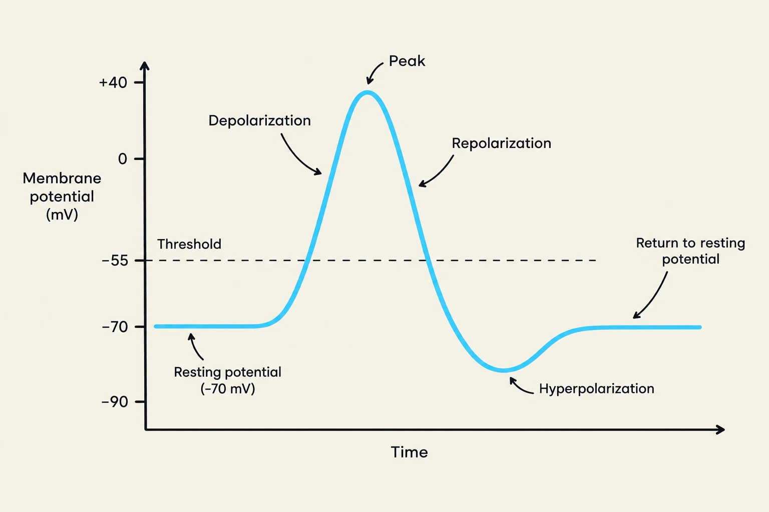
A graph illustrating the changes in membrane potential during an action potential, showing depolarization, peak, repolarization, hyperpolarization, and return to resting potential.
Once the threshold is met, an action potential (or nerve impulse) is generated. This is a rapid sequence of voltage changes that travels along the axon. The process follows a specific cycle:
1. Depolarization
When the threshold is reached, voltage-gated Na+ channels open rapidly. Because of the electrochemical gradient, Na+ floods into the axon. This influx of positive charge makes the inside of the cell less negative (moving toward zero and even becoming positive), a process called depolarization. This creates a positive feedback loop: more Na+ entry causes even more voltage-gated channels to open.
2. Repolarization
As the membrane potential peaks, voltage-gated Na+ channels inactivate, and voltage-gated $K^+$ channels open. $K^+$ ions are driven outward of the cell due to their electrochemical gradient. This loss of positive charge restores the negative internal environment, a process known as repolarization.
3. Hyperpolarization
Often, the $K^+$ channels remain open slightly longer than necessary to reach the resting state, causing the membrane potential to become even more negative than −70 mV. This phase is called hyperpolarization. Eventually, the sodium-potassium pumps and leak channels restore the membrane to its original RMP.
Refractory Periods
During an action potential, the neuron undergoes two distinct refractory periods that prevent the signal from traveling backward and limit the frequency of firing:
Absolute Refractory Period: During this time, the membrane cannot respond to any further stimulation, regardless of the strength.
Relative Refractory Period: The membrane can respond, but only to a stimulus that is larger than a normal stimulus.
Propagation of the Impulse
The action potential must travel from the soma to the synapse. The method of travel depends on the structure of the axon:
Continuous Propagation: Occurs in unmyelinated axons, where the impulse moves steadily along the entire length of the membrane.
Saltatory Propagation: Occurs in myelinated axons. The myelin sheath acts as insulation, and the action potential "jumps" from one Node of Ranvier (the gaps in the myelin) to the next. This is significantly faster than continuous propagation.
Synaptic Transmission
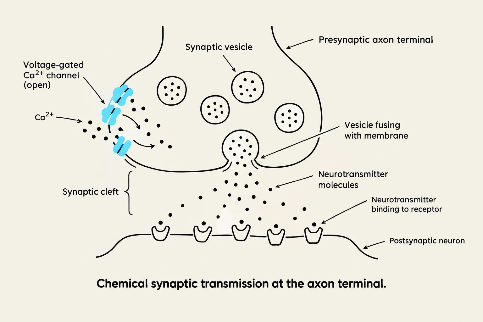
Diagram illustrating chemical synaptic transmission at the axon terminal, showing the release of neurotransmitters from the presynaptic neuron and their binding to receptors on the postsynaptic neuron.
When the impulse reaches the end of the axon, it must cross the gap between neurons, known as the synaptic cleft. There are two main types of synapses: electrical synapses and chemical synapses. In a chemical synapse, the signal is converted from electrical to chemical and back to electrical.
The Role of Neurotransmitters
The process of chemical transmission involves several steps:
Arrival of Action Potential: The impulse reaches the presynaptic axon terminal.
Calcium Influx: Depolarization opens voltage-gated calcium (Ca2+) channels. Ca2+ enters the cytosol of the terminal.
Exocytosis: The rise in calcium triggers synaptic vesicles to fuse with the membrane, releasing neurotransmitters into the synaptic cleft via exocytosis.
Binding: Neurotransmitters bind to receptors on the postsynaptic neuron.
Acetylcholine (ACh) is a primary example of a neurotransmitter. Depending on the receptor it binds to, it can cause:
Excitatory effects: Leading to depolarization and a new action potential.
Inhibitory effects: Leading to hyperpolarization, making the neuron less likely to fire.
To prevent constant stimulation, the neurotransmitter must be removed. In the case of ACh, the enzyme acetylcholinesterase (AChE) breaks it down into acetate and choline. The axon terminal then reabsorbs the choline to resynthesize more ACh.
Clinical Correlation: Various substances can alter this process. For example, Botulinum toxin prevents the exocytosis of neurotransmitters, while Novocain blocks voltage-gated sodium channels to prevent pain signals from being sent.
Keep reading for free.
Create an account to finish this reading and make it part of your study routine.