Myoglobin and Hemoglobin: Structure, Function, and Oxygen Dynamics
Explores the structure, function, and oxygen-binding dynamics of myoglobin and hemoglobin, including their heme groups, cooperativity, allosteric regulation (Bohr effect), and clinical significance.
In the complex landscape of biological respiration, the ability to capture, transport, and release oxygen is a fundamental requirement for life. This process is governed by two specialized proteins: myoglobin (Mb) and hemoglobin (Hb). While they share a common chemical foundation, their structural differences allow them to perform vastly different physiological roles—one acting as a local reservoir in muscle tissue, and the other as a sophisticated, cooperative transporter in the bloodstream.
The Heme Group and Coordination Chemistry
At the heart of both proteins lies the heme group, a prosthetic group that facilitates oxygen binding. The heme structure consists of a porphyrin ring with a central iron atom in the ferrous state, Fe(II).
The coordination of this iron atom is highly specific:
Four nitrogen atoms from the porphyrin ring coordinate the Fe(II) in a planar arrangement.
A fifth ligand, provided by the amino acid His F8 (a histidine residue located on the F helix), coordinates the iron from below the plane.
The sixth coordination site is reserved for the ligand, such as oxygen (O2).
A critical biological challenge is preventing the irreversible oxidation of Fe(II) to Fe(III) (methemoglobin), which cannot bind oxygen. The surrounding globin protein provides a protective environment that enables reversible O2 binding.
Warning: Carbon Monoxide Poisoning
Carbon monoxide (CO) is a dangerous competitor for the heme site. It binds to the Fe(II) with an affinity approximately 200 times stronger than that of oxygen, effectively blocking oxygen transport and leading to cellular hypoxia.
Myoglobin vs. Hemoglobin: Function and Structure
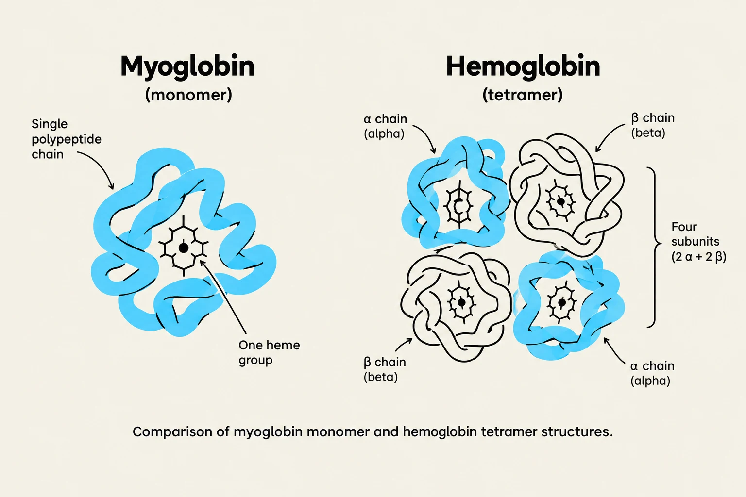
Comparison of myoglobin monomer and hemoglobin tetramer structures.
While their active sites are similar, their quaternary structures dictate their distinct roles in the body.
Myoglobin: The Oxygen Reservoir
Myoglobin is a monomer (a single polypeptide chain). Because it lacks multiple subunits, it cannot exhibit cooperativity. Its primary function is to facilitate respiration in rapidly respiring muscle tissue by increasing the solubility of oxygen and providing a local storage supply.
Hemoglobin: The Cooperative Transporter
Hemoglobin is a tetramer (α2β2), consisting of four subunits. This multi-subunit structure is essential for its role in transporting oxygen from the lungs to the tissues. Unlike myoglobin, hemoglobin's affinity for oxygen changes dynamically depending on how many oxygen molecules are already bound.
Cooperativity and the Hill Coefficient
One of the most remarkable features of hemoglobin is cooperativity. When one oxygen molecule binds to one of the four heme sites, it induces a conformational change that increases the affinity of the remaining sites for oxygen.
This phenomenon is mathematically described by the Hill coefficient (n):
A higher Hill coefficient indicates a higher degree of cooperativity.
For hemoglobin, the coefficient is typically around n=3.
This cooperativity results in a sigmoidal (S-shaped) binding curve, which allows hemoglobin to be nearly saturated in the oxygen-rich environment of the lungs while being able to release oxygen efficiently in the oxygen-poor environment of the tissues.
The T-State and R-State Transition
Diagram illustrating the structural transition between the T-state (low affinity) and R-state (high affinity) of hemoglobin, showing the movement of the iron atom within the heme group upon oxygen binding.
The transition between high and low oxygen affinity is driven by a shift between two quaternary states:
T-state (Tense): The deoxy form of hemoglobin. In this state, the protein has a lower affinity for O2. Structurally, the Fe atom sits approximately 0.6A˚ out of the heme plane.
R-state (Relaxed): The oxy form of hemoglobin. In this state, the affinity for O2 is much higher. The binding of oxygen pulls the Fe atom into the plane of the heme ring, triggering a shift in the F8 helix and a global change in the protein's quaternary structure.
Allosteric Regulation: The Bohr Effect and Beyond
To ensure oxygen is delivered exactly where it is needed most (such as metabolically active, acidic tissues), hemoglobin is regulated by several allosteric effectors that stabilize the T-state, thereby promoting oxygen release.
The Bohr Effect
The Bohr effect describes how pH influences oxygen affinity. In actively respiring tissues, CO2 production leads to an increase in hydrogen ion concentration ([H+]), lowering the pH.
Lower pH (Higher [H+]): Promotes the T-state, facilitating the release of O2.
Carbon dioxide is transported in the blood via two main routes:
Bicarbonate Pathway: The majority of CO2 is converted into bicarbonate (HCO3−) by the enzyme carbonic anhydrase.
Carbamate Formation:CO2 can also react directly with the N-termini of the hemoglobin subunits to form carbamates. This reaction produces $H^+$ (contributing to the Bohr effect) and helps stabilize the T-state.
2,3-bisphosphoglycerate (BPG)
2,3-BPG is a small molecule that binds to the central cavity of the hemoglobin tetramer, specifically stabilizing the T-state. By decreasing hemoglobin's affinity for oxygen, BPG ensures efficient unloading in the tissues. An increase in BPG concentration is a critical adaptive response for humans living at high altitudes, where oxygen pressure is lower.
Clinical Significance: Hemoglobin Mutants
Because the function of hemoglobin depends so precisely on its structure, even a single amino acid substitution can have devastating consequences. There are approximately 500 known variants of hemoglobin.
A classic example is Sickle Cell Anemia. In this condition, a mutation leads to the formation of S-fibers (aggregates of hemoglobin). These fibers distort the red blood cell into a characteristic "sickle" shape, leading to sickled erythrocytes that can clog capillaries and impede blood flow.
Keep reading for free.
Create an account to finish this reading and make it part of your study routine.