


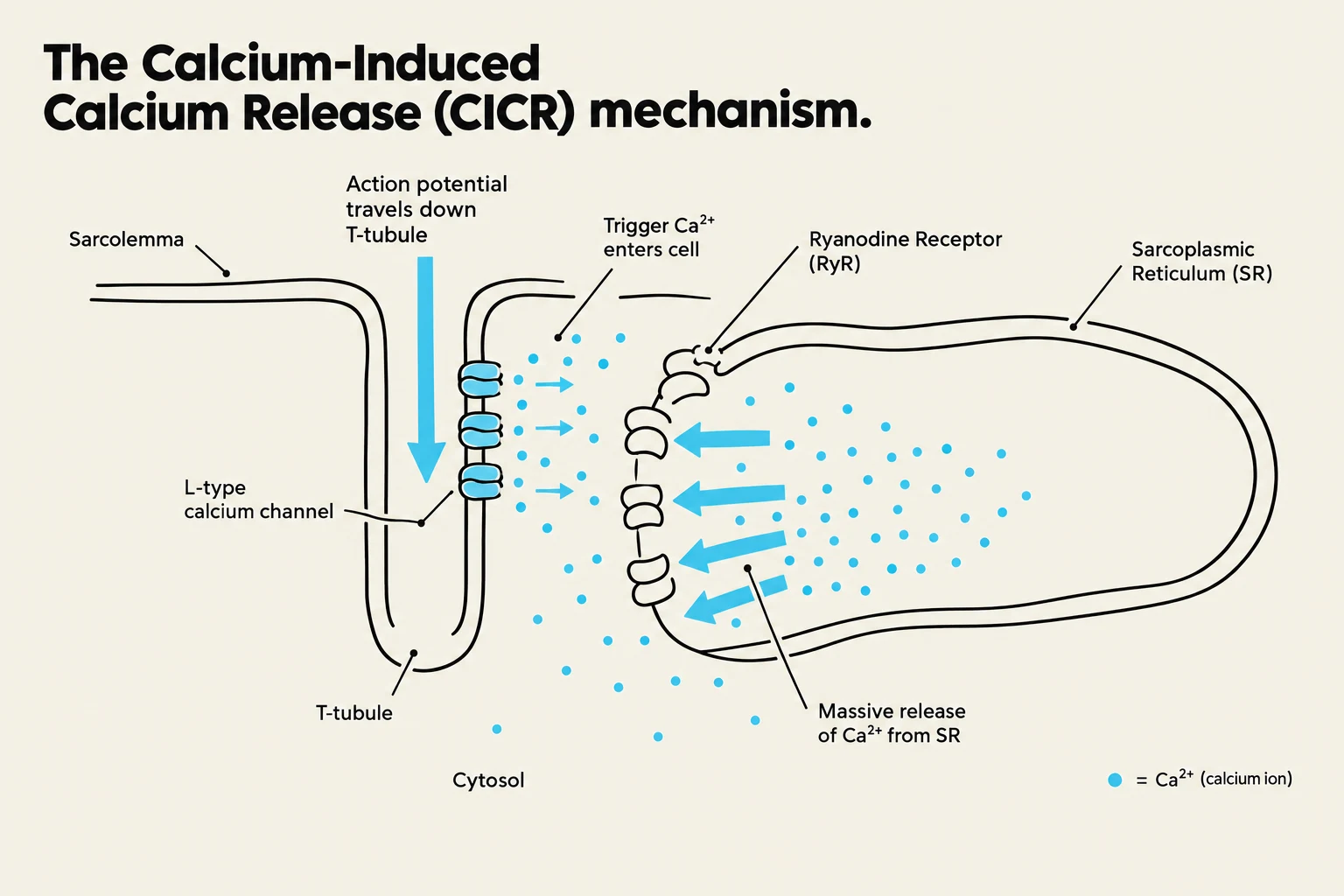
To understand how the heart functions as a pump, one must look beyond the organ level and zoom into the microscopic architecture of the individual cardiac muscle cell, or myocyte. The heart does not simply contract through random electrical impulses; it relies on a highly coordinated sequence where electrical signals are converted into mechanical force. This process is known as Excitation-Contraction Coupling (ECC).
In this article, we will explore the specialized structural components that allow myocytes to communicate and contract, and the intricate calcium signaling pathways that drive the heartbeat.
The Microanatomy of the Myocyte
T-Tubules: The Electrical Highways
Because cardiac myocytes are relatively large in diameter, an electrical impulse traveling only along the surface membrane (the sarcolemma) would take too long to reach the deep interior of the cell. To solve this, the sarcolemma invaginates deep into the cell, forming a network of small tubes known as Transverse Tubules, or T-tubules.
These T-tubules ensure that the action potential is delivered almost simultaneously to the entire volume of the cell. This rapid depolarization is essential for ensuring that all myofibrils (the contractile proteins) within the cell contract at once, rather than in a staggered, inefficient wave.
Intercalated Discs: Ensuring Syncytium
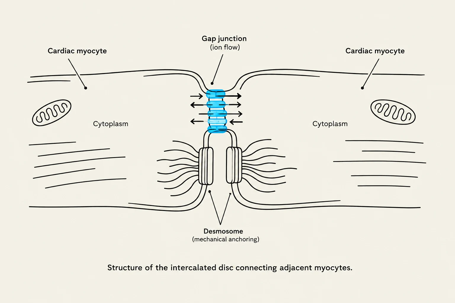
Unlike skeletal muscle, where fibers act independently, cardiac muscle cells must act as a single functional unit, often referred to as a functional syncytium. This is made possible by intercalated discs, the specialized junctions that connect adjacent myocytes.

Keep reading for free.
Create an account to finish this reading and make it part of your study routine.
Continue reading freeAlready have an account? Log in
Updated