Myeloproliferative Disorders and Polycythemia Vera
An overview of myeloproliferative disorders (MPDs), including Polycythemia Vera (PV) and Essential Thrombocythemia (ET). Discusses their causes, laboratory findings, clinical consequences, and treatment strategies.
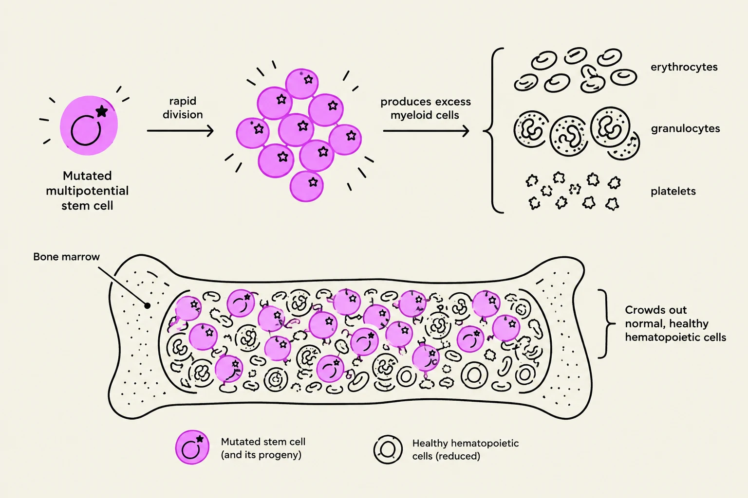
Hematologic malignancies are broadly categorized into leukemias and myeloproliferative disorders (MPDs). While both involve the malignant transformation of hematopoietic cells, they differ in their clinical presentation and cellular characteristics. Myeloproliferative disorders are clonal hematopoietic stem cell diseases characterized by the expansion, excessive production, and over-accumulation of one or more myeloid cell lines, including erythrocytes, granulocytes, and platelets. These conditions are often considered "pre-leukemic" because they can eventually transition into acute leukemias.
Understanding Myeloproliferative Disorders (MPDs)
This diagram illustrates how a mutated multipotential stem cell in the bone marrow undergoes rapid division, producing excess myeloid cells. These mutated cells crowd out normal, healthy hematopoietic cells, leading to a reduction in erythrocytes, granulocytes, and platelets.
MPDs arise from a mutation in a single multipotential or pluripotential stem cell. This mutation causes the cell to proliferate abnormally, eventually taking over the bone marrow and preventing the production of normal cells. These disorders are typically chronic in nature, though they may progress through accelerated, subacute, or acute phases.
Common clinical symptoms of MPDs are often insidious, developing slowly and appearing vague. Patients may present with:
Fatigue and anorexia
Splenomegaly (enlarged spleen)
Bone pain due to marrow expansion
The disease course can vary significantly; while some patients live for years, others may succumb to complications such as infection, hemorrhage, or thrombosis.
Polycythemia Vera (PV)
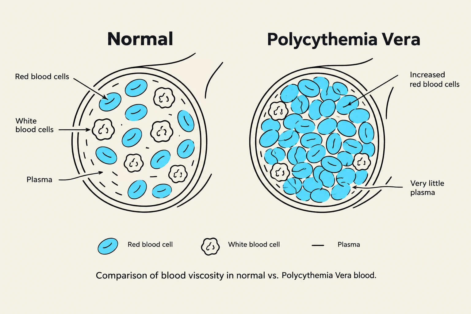
Comparison of blood viscosity in normal vs. Polycythemia Vera blood.
Polycythemia Vera is a specific stem cell disorder that affects all three myeloid cell lines: red blood cells (RBCs), white blood cells (WBCs), and platelets. Although all lines are increased, the RBCs are the dominant cell type affected, leading to a significant increase in total RBC mass and blood volume.
Laboratory Findings in PV
Diagnosis of PV is supported by specific laboratory markers:
Hemoglobin levels: Typically >18.5 gm/dL in males and >16.5 gm/dL in females.
Peripheral Blood: May show a "left shift" in WBCs and the presence of occasional nucleated RBCs (NRBCs).
Clinical Consequences and Treatment
The primary clinical danger in PV is increased blood viscosity caused by the excessive red cell mass. This high viscosity significantly raises the risk of life-threatening events, including thrombosis, stroke, and heart attack. Over time, the disorder may progress to myelofibrosis, where the bone marrow becomes fibrotic.
Treatment strategies for PV include:
Therapeutic phlebotomy (to reduce blood volume).
Myelosuppressive drugs.
Radioactive phosphorus.
💡 Exam Tip: Distinguishing Polycythemias
It is vital to distinguish PV from other forms of polycythemia:
Relative Polycythemia: Caused by a decrease in plasma volume (e.g., dehydration, obesity, or heavy smoking), making the blood appear concentrated.
Secondary Polycythemia: Caused by increased erythropoietin or decreased oxygen delivery (e.g., COPD or emphysema).
Polycythemia Vera: A primary clonal disorder with decreased EPO levels.
Essential Thrombocythemia (ET)
This illustration compares normal platelets with those found in Essential Thrombocythemia, highlighting morphological differences and including a micromegakaryocyte.
Essential Thrombocythemia is a clonal disorder involving a single multipotential stem cell that leads to the excessive production of platelets.
Laboratory and Morphological Hallmarks
Platelet Count: Extremely high, often >1,000,000/uL.
Morphology: Peripheral blood may show "huge and bizarre" platelet forms, as well as micromegakaryocytes and bare nuclei.
Other findings: An increase in WBCs with a left shift and normocytic, normochromic (NC/NC) anemia are common.
Clinical Presentation and Treatment
While ET is characterized by high platelet counts, these platelets are often easily activated, leading to a high risk of thrombosis (clots) and vascular problems. Conversely, because the platelets may malfunction, patients can also suffer from hemorrhage. Treatment typically involves the use of immunosuppressive drugs or radioactive phosphorus.
🔍 Comparison: ET vs. Relative Thrombocytosis
Essential Thrombocythemia (ET): A clonal disorder where platelets are often morphologically abnormal (huge/bizarre).
Relative Thrombocytosis: A non-clonal increase in platelets caused by external factors like iron deficiency, infection, hemorrhage, or malignancy. In these cases, the platelets themselves appear morphologically normal.
Summary of Myeloproliferative Disorders
Disorder
Primary Cell Line Affected
Key Feature
Polycythemia Vera
Erythrocytes (RBCs)
Increased viscosity; Low EPO
Essential Thrombocythemia
Platelets
Huge/bizarre platelets; High risk of thrombosis
Primary Myelofibrosis
Megakaryocytes/All lines
Bone marrow fibrosis; Dacrocytes (tear-drop cells)
Keep reading for free.
Create an account to finish this reading and make it part of your study routine.