Explore the agents that cause DNA mutations, the types of damage they inflict, and the cellular repair mechanisms that protect the genetic blueprint. Understand how mutations impact gene function and are used in genetic research.
In the study of genetics, mutations are far more than just "errors." They are powerful tools that allow scientists to probe the very essence of life. By observing how a change in a DNA sequence alters a protein, researchers can deduce the original function of a gene. To understand how these changes occur and how cells fight to prevent them, we must look at the chemical and physical forces that attack our genetic blueprint, the damage they cause, and the sophisticated repair machinery that keeps us alive.
The Nature of Mutagens
A mutagen is any agent—chemical, physical, or biological—that increases the frequency of mutations by damaging DNA. These agents can be categorized based on how they interact with the double helix.
Physical Mutagens: Radiation
Radiation is one of the most potent sources of DNA damage. It is generally divided into two types:
Ionizing Radiation: High-energy waves, such as X-rays, can physically break the sugar-phosphate backbone of the DNA, leading to double-strand breaks.
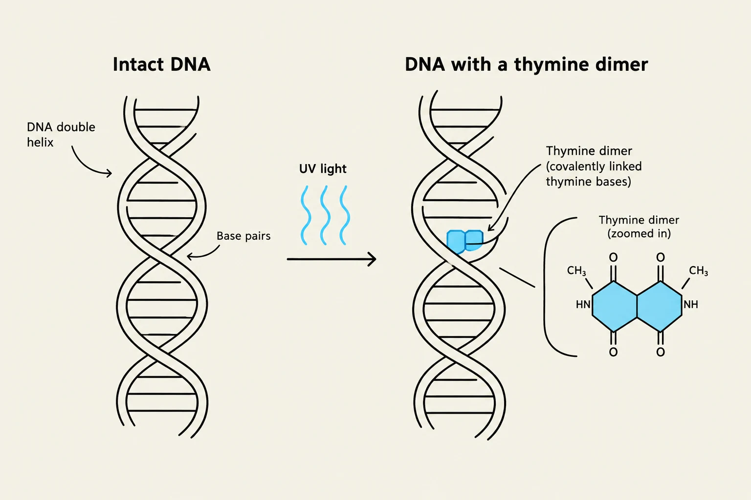
UV Radiation: Ultraviolet light (sunlight) does not typically break the backbone but instead causes adjacent pyrimidine bases (usually two thymines) to bond together covalently, forming thymine dimers.
Illustration showing how UV light can cause adjacent thymine bases in DNA to form a thymine dimer, a type of DNA damage.
Chemical Mutagens
Chemical agents can alter DNA in several subtle, yet devastating, ways:
Deaminating Agents: These remove amino groups from bases. For example, nitrous acid can convert cytosine into uracil, which changes how the base pairs during replication.
Alkylating Agents: Compounds like EMS or MMS add methyl or ethyl groups to bases, altering their ability to pair correctly.
Base Analogs: These are chemicals that "mimic" normal bases. Because they look so much like the standard nucleotides, the cell mistakenly incorporates them into the DNA during replication.
Intercalating Agents: Molecules like ethidium bromide wedge themselves between the stacked bases of the DNA helix. This distorts the shape of the helix and often leads to frameshift mutations.
Cellular Defense: DNA Repair Mechanisms
Because DNA is constantly under attack, cells have evolved a specialized toolkit of repair mechanisms. The specific mechanism used depends entirely on the type of damage encountered.
Single-Base and Small Lesion Repair
When a single base is damaged or incorrectly paired, the cell employs specialized pathways:
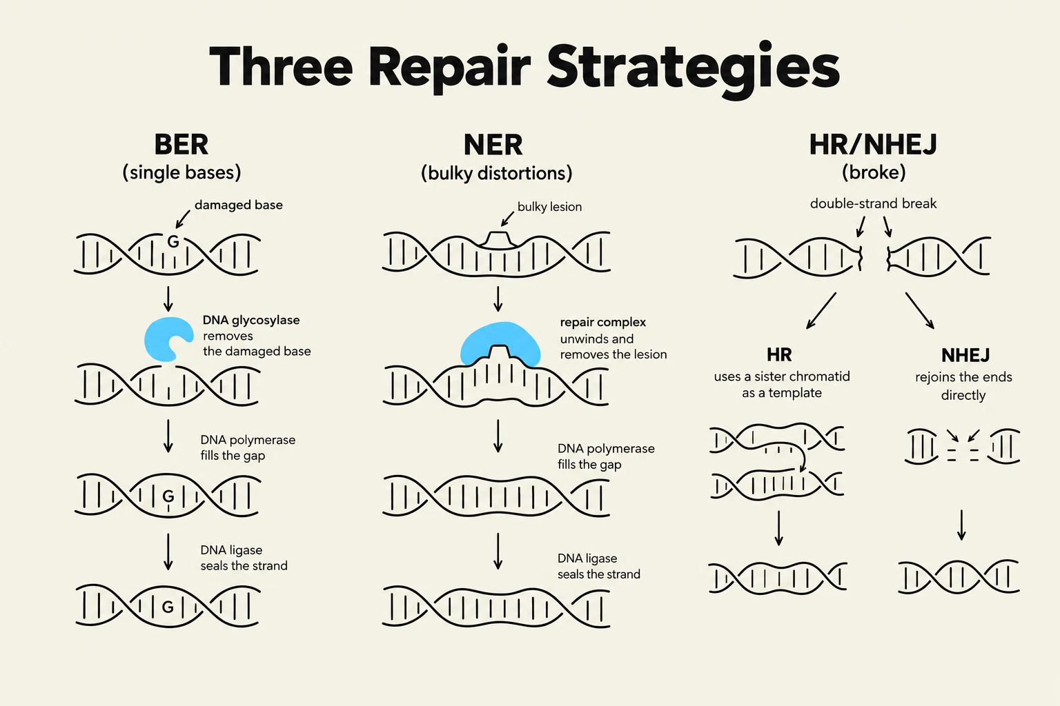
Base Excision Repair (BER): This is the primary defense against deamination (e.g., cytosine to uracil) and alkylation. It involves removing the damaged base and replacing it with the correct one.
Mismatch Repair (MMR): This system acts as a "spell-checker" during or immediately after replication to correct base mismatches or small insertions/deletions that escaped the DNA polymerase.
Structural and Helix Distortion Repair
When the damage is more extensive and affects the physical structure of the DNA strand, other pathways take over:
Nucleotide Excision Repair (NER): This mechanism is crucial for fixing bulky lesions that distort the double helix, such as the thymine dimers caused by UV light or the distortions caused by intercalating agents.
Repairing Double-Strand Breaks
If a chromosome is severed entirely (a double-strand break), the cell faces a crisis. It has two main options:
Homologous Recombination (HR): A high-fidelity process that uses a sister chromatid as a template to perfectly reconstruct the broken sequence.
Non-Homologous End Joining (NHEJ): A faster but more "error-prone" method where the cell simply glues the broken ends back together, which often results in the loss or addition of a few nucleotides.
Diagram illustrating three DNA repair strategies: Base Excision Repair (BER), Nucleotide Excision Repair (NER), and Homologous Recombination/Non-Homologous End Joining (HR/NHEJ).
From Mutation to Function: The Polypeptide Chain
To understand why these repairs are so critical, we must look at the end product: the polypeptide chain. Genes specify the identity and order of amino acids in a protein. Because the three-dimensional shape and function of a protein are determined by this specific sequence, even a single mutation can have catastrophic effects.
Types of Mutations in the Genetic Code
The genetic code is a triplet code, meaning three nucleotides (a codon) specify one amino acid. Mutations can disrupt this in three primary ways:
Missense Mutations: A single nucleotide change results in the substitution of one amino acid for another. A classic example is Sickle Cell Disease, where a single amino acid substitution (Glu → Val) in the hemoglobin β chain causes the protein to aggregate, distorting red blood cells into a sickle shape.
Nonsense Mutations: A mutation creates a premature stop codon (UAA, UAG, or UGA), which halts translation mid-way through the polypeptide chain, usually resulting in a non-functional, truncated protein.
Frameshift Mutations: Caused by the insertion or deletion of nucleotides (that are not in multiples of three), these mutations shift the entire reading frame. This changes every single amino acid downstream of the mutation, completely altering the protein's identity.
Exam Tip: Remember that the genetic code is degenerate, meaning multiple codons can code for the same amino acid, but it is unambiguous, meaning one specific codon only codes for one specific amino acid. This degeneracy can sometimes buffer the effects of certain mutations!
Using Mutations to Map Genes
Scientists don't just study mutations to see what goes wrong; they use them to map how biological systems work.
Complementation Tests
If an organism has two different recessive mutations, we can perform a complementation test to see if those mutations are in the same gene or different genes.
No Complementation: If the offspring shows the mutant phenotype, the two mutations are in the same gene.
Complementation: If the offspring shows the wild-type phenotype, the mutations are in different genes.
Supplementation Tests
By observing which nutrients a mutant can no longer produce (auxotrophs), and then "supplementing" the medium with specific chemicals, researchers can determine the order of enzymes in a metabolic pathway. If adding a specific chemical allows a mutant to grow, that chemical is likely the substrate for the enzyme that the mutant is missing.
Keep reading for free.
Create an account to finish this reading and make it part of your study routine.