


Effective management of musculoskeletal disorders requires a combination of vigilant clinical assessment and the precise application of diagnostic technologies. Whether a patient presents with a simple sprain or a complex fracture, the clinician's ability to monitor neurovascular integrity and interpret imaging results is fundamental to preventing long-term complications like compartment syndrome or permanent nerve damage.
Neurovascular Assessment: The 7 Ps
The most critical immediate assessment in musculoskeletal care is the neurovascular check. This is used to detect complications such as vascular compromise or compartment syndrome. Nurses often utilize the "7 Ps" framework to ensure a systematic evaluation of the affected extremity.
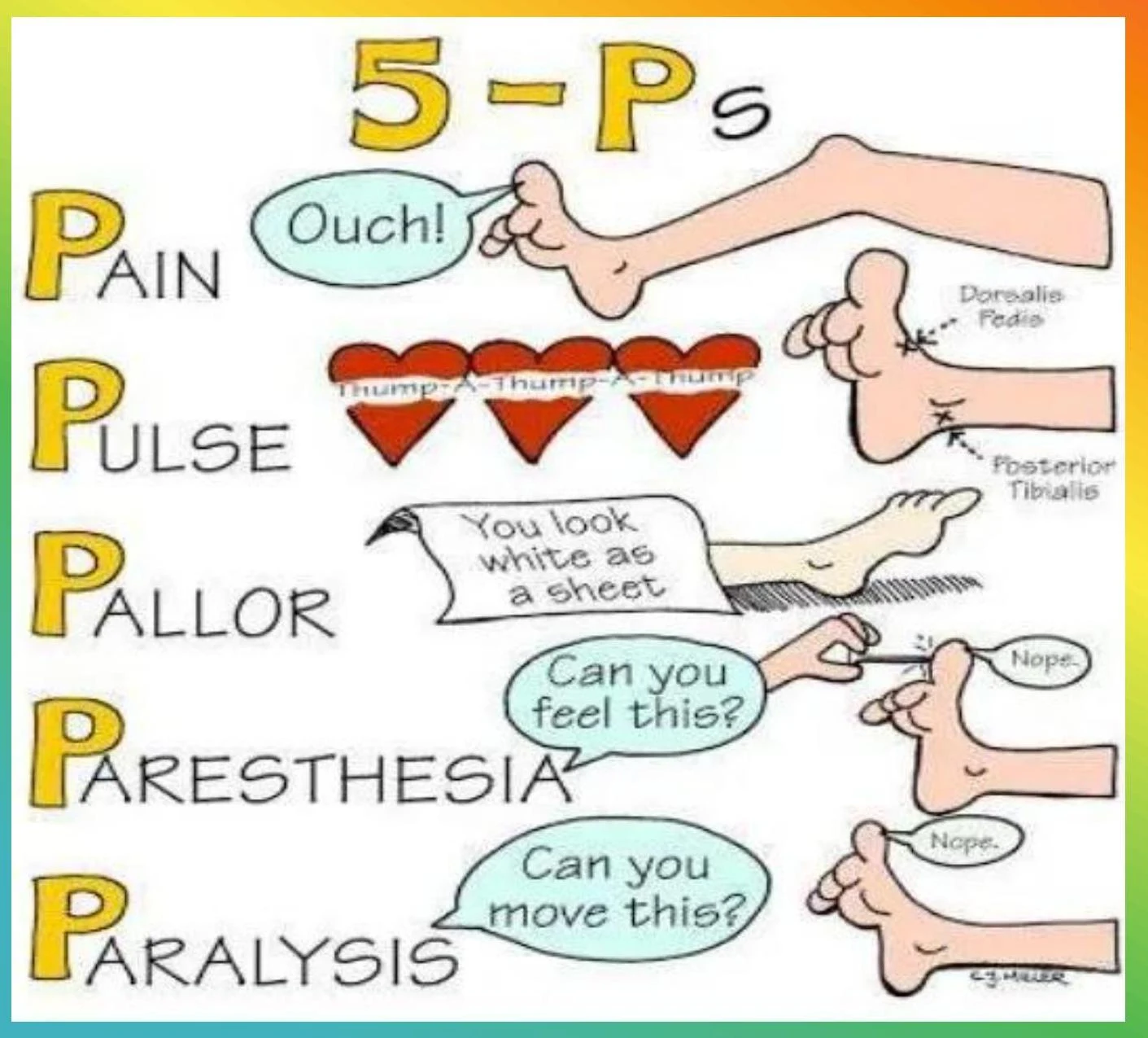
The 7 Ps include:
- Pain: Often reported as being disproportionate or worse than expected for the injury.
- Pallor: Paleness of the skin, indicating potential circulatory issues.
- Paresthesia: A "pins and needles" sensation or a loss of sensation.
- Paralysis: The inability to move the extremity.
- Pulselessness: The absence of palpable pulses in the distal extremity.
- Polar: The extremity feels cold to the touch.
- Puffiness: Swelling or edema in the area.
Clinical Tip: If a patient reports pain that is significantly worse than the injury would suggest, or if they experience sudden numbness, notify the provider immediately, as these can be early signs of compartment syndrome.
Diagnostic Imaging: MRI and CT Scans
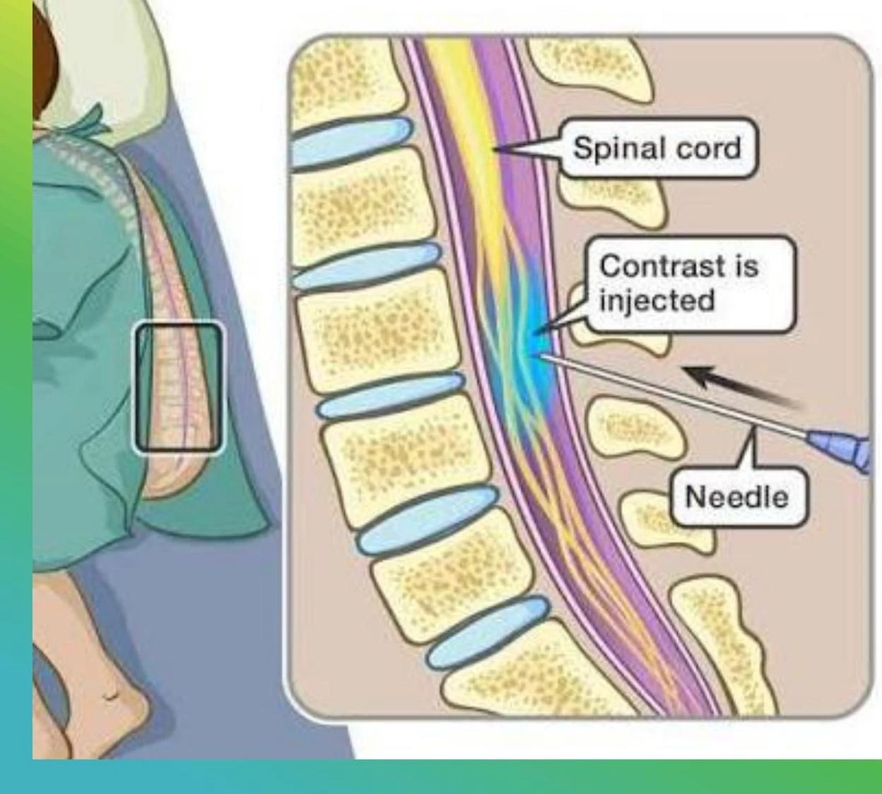
When structural damage is suspected, imaging is used to visualize internal tissues and bone architecture.
Keep reading for free.
Create an account to finish this reading and make it part of your study routine.
Continue reading freeAlready have an account? Log in
Updated