A review of muscle anatomy, motor units, and various muscle conditions including primary myopathies, inflammatory conditions, and general muscle problems.
The muscular system is a complex network of tissues designed for movement, stability, and heat production. Understanding how muscles function requires a look at their microscopic architecture, the neurological signals that trigger them, and the various ways these systems can fail due to genetics, inflammation, or trauma.
Muscle Anatomy and Function
At the most fundamental level, the structural unit of skeletal muscle is the muscle fiber. These fibers do not exist in isolation; each level of muscle organization is enveloped by layers of connective tissue. This connective tissue is vital because it houses the nerves, blood vessels, and lymphatic vessels necessary to supply the muscle with nutrients and regulatory signals.
To achieve coordinated movement, the nervous system utilizes the motor unit. A motor unit consists of a single motor neuron and all the muscle fibers that the neuron stimulates. These units are recruited asynchronously, meaning they do not all fire at once. This staggered recruitment is a critical physiological mechanism that delays muscle fatigue and allows for smooth, sustained muscular contractions.
Diagram illustrating a motor neuron connecting to multiple muscle fibers, explaining how a single signal can activate many fibers simultaneously.
Muscles also undergo physical changes in response to their environment:
Hypertrophy: An increase in the size of tissue or organ caused by the enlargement of individual cells, often seen in skeletal or cardiac muscle due to increased workload.
Atrophy: The wasting of normally developed tissue due to cell degeneration, which can result from malnourishment, disuse (such as immobilization), aging, or denervation.
General Muscle Problems and Abnormalities
Muscle dysfunction can manifest as a wide array of involuntary movements or physical sensations. These range from minor twitches to violent, systemic contractions.
Involuntary Movements and Spasms
Cramps (Spasms): These are prolonged, painful, and involuntary contractions of a specific muscle or group. They can be idiopathic or linked to metabolic diseases and electrolyte depletion, particularly involving sodium (Na+), potassium ($K^+$), or calcium (Ca2+).
Fasciculation: A brief, involuntary contraction of a single motor unit, often visible as a "flicker" under the skin.
Fibrillation: Unlike fasciculation, this is the spontaneous, rhythmical contraction of a single muscle fiber.
Tetany: A state of increased neuromuscular excitability characterized by spasms and twitching, often caused by electrolyte imbalances (such as low Ca2+ or Mg2+) that cause unstable depolarization of motor neurons.
Myoclonus: Sudden, unexpected, brief contractions of limb muscles, which can be benign (associated with sleep) or pathological (associated with seizures or cerebral hypoxia).
Convulsion: A sudden, violent, involuntary contraction of muscle groups that produces contortions of the body, such as in febrile seizures.
Physical Stress and Structural Damage
Strain: An injury involving the excessive stretching or partial tearing of a muscle or tendon.
Contracture: A condition where muscle fibers shorten without an action potential, often caused by a failure of the calcium pump in the sarcoplasmic reticulum.
Stress-induced muscle tension: Often associated with chronic anxiety, this manifests as neck stiffness, back pain, or clenching of the teeth.
Exam Tip: Distinguish between a strain and a sprain. A strain refers to injury to a muscle or tendon, whereas a sprain refers to an injury to a ligament.
Primary Myopathies
Primary myopathies are diseases that are intrinsic to the muscle fiber itself, often resulting from heritable or autoimmune factors.
Muscular Dystrophy
Muscular dystrophies are genetic disorders characterized by the progressive degeneration and necrosis of skeletal muscle fibers. These are often caused by mutations in the dystrophin gene. Dystrophin is a protein that normally anchors the muscle cell cytoskeleton to the extracellular matrix (ECM), protecting the cell from mechanical damage during contraction.
Two major forms include:
Duchenne Muscular Dystrophy (DMD): The most common form, caused by a complete lack of dystrophin due to a frameshift mutation. It typically results in a clumsy gait in infancy and often leads to wheelchair use by age 12.
Becker Muscular Dystrophy: A milder form where patients possess an abnormal but semi-functional dystrophin protein, leading to a slower disease progression.
Myotonic Dystrophy
This is a group of autosomal dominant disorders distinguished by myotonia—the inability of the muscle to relax after a contraction. It is caused by repeat expansions in specific genes (like DMPK or CNBP) that lead to abnormal mRNA clumps, interfering with the expression of other essential genes.
Inflammatory and Autoimmune Conditions
Some muscle problems arise not from the muscle fiber's DNA, but from the body's immune system attacking the neuromuscular junction or the muscle tissue itself.
Myasthenia Gravis
Myasthenia gravis is a chronic autoimmune disorder that impairs communication between nerves and muscles. Auto-antibodies attack the acetylcholine (Ach) receptors at the neuromuscular junction (NMJ). This reduces the number of functional receptors, leading to decreased depolarization and muscle weakness. A hallmark of this condition is fatigue that worsens with activity and improves with rest.
Fibromyalgia Syndrome (FMS)
FMS is a chronic pain condition characterized by diffuse, achy musculoskeletal pain, fatigue, and sleep disturbances. Unlike many other conditions, FMS shows no evidence of tissue destruction or inflammation in laboratory tests; it is thought to be related to CNS disturbances that increase pain perception.
Rhabdomyolysis
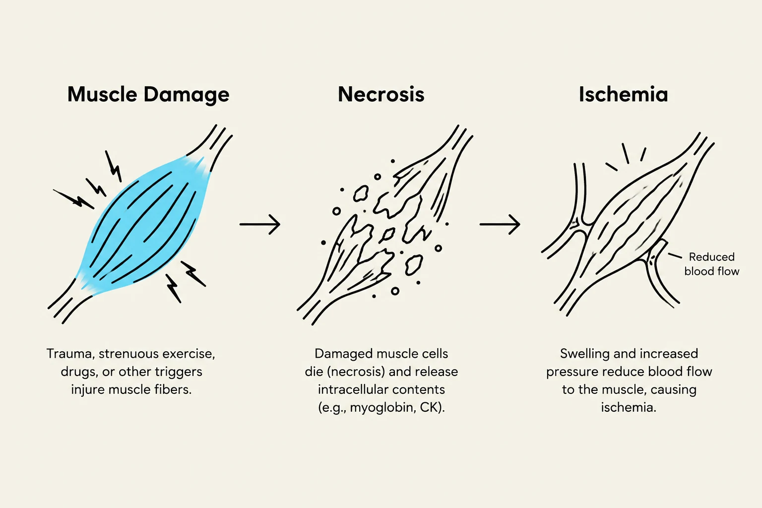
Rhabdomyolysis is a life-threatening condition involving severe muscle trauma and muscle cell loss. When muscle cells are damaged (due to crush syndrome, compartment syndrome, or certain medications), they undergo necrosis. This releases myoglobin into the bloodstream (hypermyoglobinemia), which can then deposit in the renal tubules, leading to kidney damage (myoglobinuria).
This diagram illustrates the progression from muscle damage to necrosis and ischemia, detailing the causes and effects at each stage.
Keep reading for free.
Create an account to finish this reading and make it part of your study routine.