Molecular cloning is a fundamental technique in genetic engineering used to isolate, amplify, and manipulate specific DNA sequences. The process involves inserting a target DNA fragment (the "insert") into a DNA molecule called a "vector" (often a plasmid) to create a recombinant product. By successfully combining these elements, scientists can transform bacteria to produce specific proteins or study gene function. This article explores the enzymatic strategies and specialized tools used to ensure efficient cloning and the creation of stable recombinant DNA.
Restriction Digestion Strategies
The first step in most cloning workflows is preparing the DNA using restriction enzymes. These enzymes act as molecular scissors, cutting DNA at specific recognition sequences. The choice between single and double digestion significantly impacts the efficiency and directionality of the cloning process.
Single Digestion
In a single digestion, only one restriction enzyme is used to cut both the vector and the insert. Because the two ends of the DNA are identical and compatible, they can easily re-anneal. While this is a simple method, it presents two major challenges:
Vector Self-Ligation: The vector can close back up on itself without incorporating the insert.
Lack of Orientation: The insert can be integrated into the vector in two different directions (forward or reverse), which may be problematic if the gene needs to be expressed under a specific promoter.
Double Digestion
To overcome these issues, researchers often use two different restriction enzymes to perform a double digestion. This creates two different "sticky ends" (incompatible overhangs) at the ends of the DNA fragments.
Using two different enzymes provides several advantages:
Directional Cloning: The insert can only fit into the vector in one specific orientation because its ends must match the corresponding ends of the vector.
Reduced Self-Ligation: Since the two ends of the vector are no longer compatible, the vector is much less likely to close without an insert.
Increased Efficiency: Double digestion can increase the overall completion of the cleavage process.
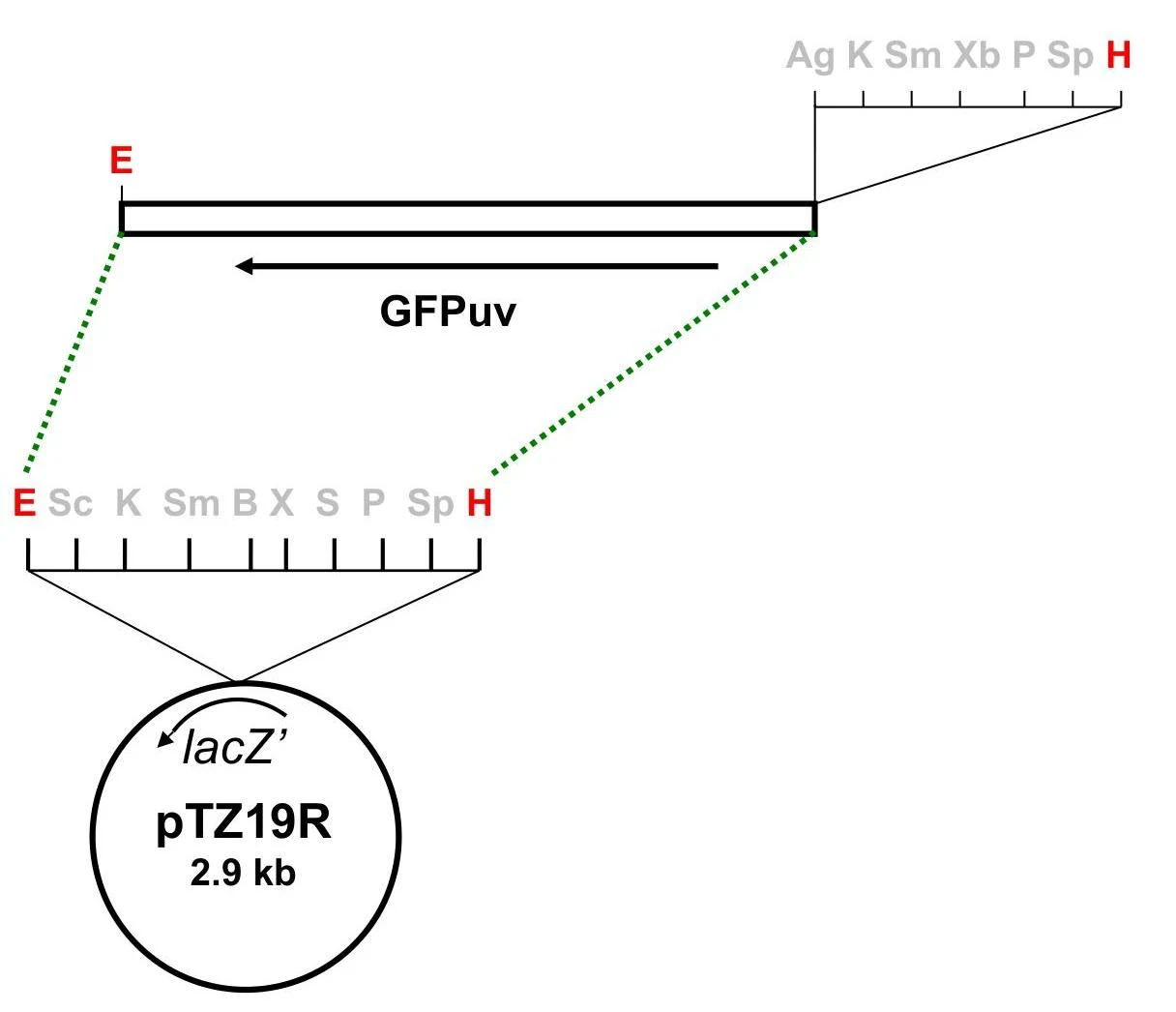
Diagram illustrating the pTZ19R plasmid with the GFPuv gene insert and restriction enzyme sites.
Diagram illustrating the pTZ19R plasmid with the GFPuv gene insert and restriction enzyme sites.
DNA Ligation and T4 DNA Ligase
Once the vector and insert have been digested, they must be physically joined. This is achieved through ligation, a process catalyzed by the enzyme T4 DNA ligase.
The Mechanism of Ligation
T4 DNA ligase facilitates the formation of a phosphodiester bond between the adjacent 3'-hydroxyl (-OH) group and the 5'-phosphate (-P) group of the DNA strands.
Note on Ligation Products: Even after successful ligation, the resulting circular DNA often contains "nicks"—breaks in the sugar-phosphate backbone where one strand is not covalently joined to the next. These nicks are typically repaired by the host bacterium's internal machinery after the DNA has been transformed into the cell.
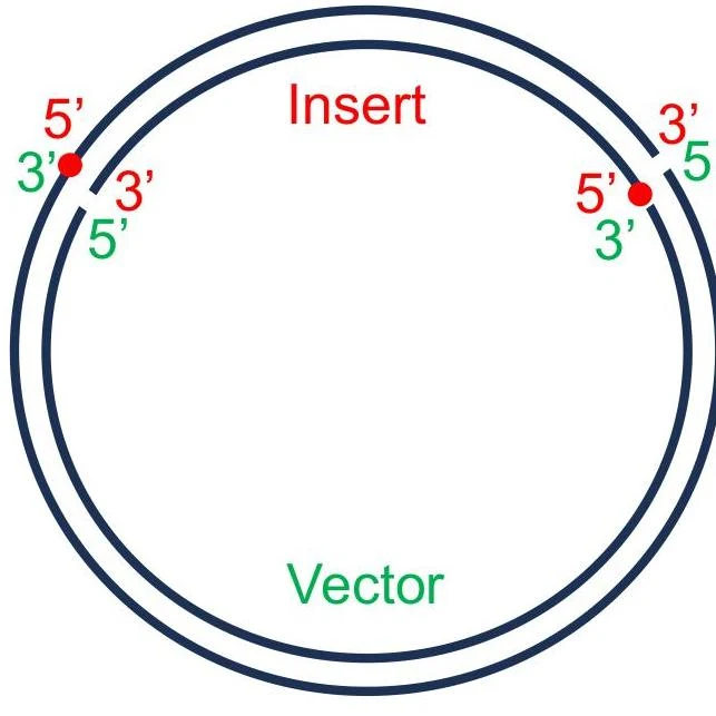
Diagram illustrating the insertion of a DNA fragment (insert) into a circular DNA molecule (vector), showing the 5' and 3' ends.
Optimizing Ligation Reactions
To maximize the yield of the desired recombinant product, several experimental parameters must be controlled:
Molar Ratios: It is standard practice to maintain an ideal Insert-to-Vector (I:V) molar ratio, typically around 3:1, though ranges from 1:1 to 10:1 may be used depending on the application.
Buffer Composition: The ligation buffer contains ATP, which is essential for the enzyme's activity. Because ATP is sensitive, the buffer should be kept on ice and subjected to minimal freeze-thaw cycles. The buffer also contains PEG, which makes the solution viscous.
Preventing Self-Ligation via Dephosphorylation: To further prevent the vector from re-ligating to itself, the 5' ends of the vector can be treated with alkaline phosphatase to remove the phosphate groups. Since T4 DNA ligase requires a 5' phosphate to form a bond, a dephosphorylated vector cannot close on itself. The insert, which still possesses its 5' phosphates, can then provide the necessary groups to bridge the gap.
Linkers and Adaptors
Sometimes, a researcher needs to clone a DNA fragment into a vector that does not have compatible restriction sites. In these cases, synthetic oligonucleotides known as linkers or adaptors can be used to "modify" the ends of the DNA.
Linkers: These are short, double-stranded DNA molecules that have two blunt ends and contain a specific restriction enzyme recognition site. Once ligated to the blunt ends of an insert, the DNA can be digested with the corresponding enzyme to create sticky ends.
Adaptors: These are more specialized molecules that contain one blunt end and one sticky end. They are particularly useful for connecting a blunt-ended fragment directly to a vector with a specific overhang without the need for further digestion.
Feature
Linker
Adaptor
Ends
Two blunt ends
One blunt, one sticky end
Requirement
Requires subsequent digestion
Can be used directly
Topoisomerase-Mediated Ligation
An alternative to the traditional T4 DNA ligase method is topoisomerase-mediated ligation, often referred to as TOPO cloning.
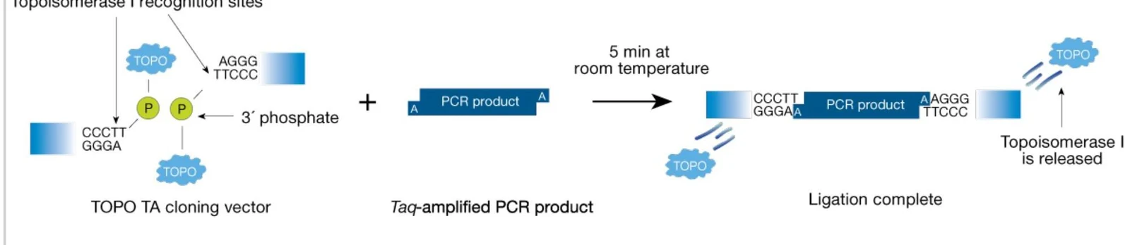
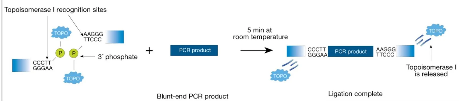
Topoisomerases are naturally occurring enzymes that regulate DNA topology by changing the supercoiling of DNA during replication and transcription. In commercial cloning kits, a specific enzyme—Vaccinia virus topoisomerase I—is covalently attached to the 3' ends of a pre-linearized vector.
When a DNA molecule (such as a PCR product) is added to this vector, the topoisomerase enzyme recognizes the DNA and facilitates the joining of the two molecules, releasing itself in the process. This method is highly valued because it is significantly faster and more efficient than standard ligation, especially for blunt-ended products.
Diagram illustrating the TOPO TA cloning process where a Taq-amplified PCR product is ligated into a TOPO TA cloning vector with the release of Topoisomerase I.
Diagram illustrating the process of ligation of a blunt-end PCR product using Topoisomerase I, showing recognition sites, the enzyme's action, and its release upon completion.
Screening Recombinant Products
After ligation and transformation into bacteria, it is necessary to identify which colonies actually contain the desired recombinant plasmid. This can be done through several screening methods:
Phenotypic Screening: Using markers like GFP (Green Fluorescent Protein). For example, on an IPTG+UV plate, one might look for white fluorescent colonies.
Blue/White Screening: Using X-gal agar plates to distinguish between colonies with and without an insert based on color changes.
Molecular Analysis: Confirming the presence and orientation of the insert via restriction digestion analysis, colony PCR, or measuring changes in molecular size through agarose gel electrophoresis.
Exam Tip: When screening, remember that a "positive" result is the presence of the recombinant product (vector + insert), while a "negative" result might be a vector that has self-ligated or a non-fluorescent colony.
Keep reading for free.
Create an account to finish this reading and make it part of your study routine.