Molecular Biology: From Genes to Proteins and Lab Techniques
Explore the fundamental processes of molecular biology, including DNA replication, transcription, and translation, which govern the flow of genetic information. Learn about key laboratory techniques like PCR and electrophoresis used to analyze biological molecules.
Molecular biology is the study of how molecules store, copy, and use biological information. This information flow is central to life, dictating how a cell functions, grows, and reproduces. By understanding the mechanisms of DNA, RNA, and proteins, we can unlock the secrets of genetics, disease, and biotechnology.
The Building Blocks: Nucleotides and Nucleic Acids
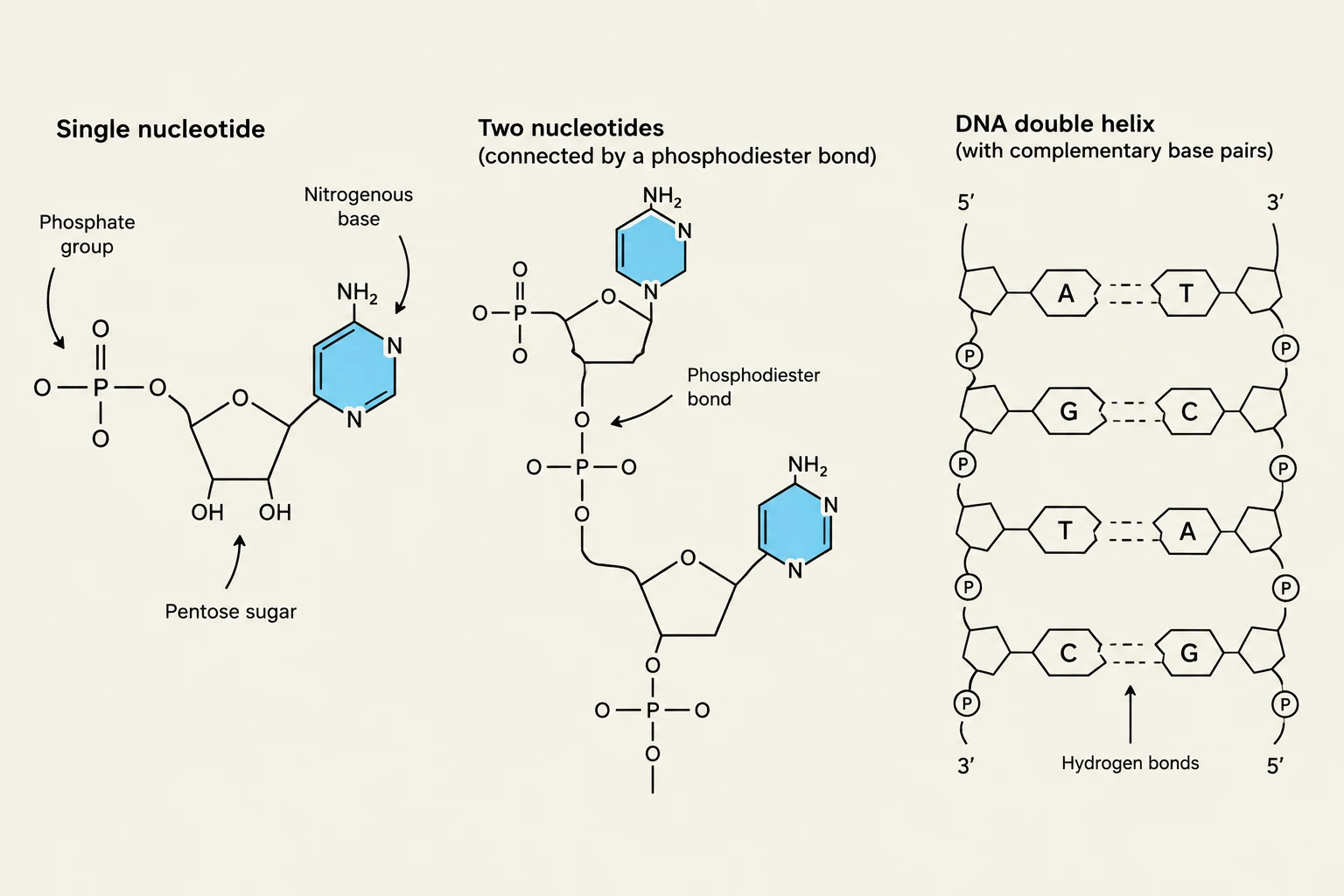
Diagrams illustrating a single nucleotide, two nucleotides connected by a phosphodiester bond, and the DNA double helix with complementary base pairs and hydrogen bonds.
At the heart of molecular biology are nucleic acids: DNA (deoxyribonucleic acid) and RNA (ribonucleic acid). These large molecules are polymers made of repeating units called nucleotides.
Every nucleotide consists of three distinct components:
A nitrogenous base
A sugar (deoxyribose in DNA; ribose in RNA)
A phosphate group
Nitrogenous Bases: Purines and Pyrimidines
The nitrogenous bases carry the actual genetic code. They are categorized into two families based on their chemical structure:
Purines (two rings): Adenine (A) and Guanine (G).
Pyrimidines (one ring): Cytosine (C), Thymine (T), and Uracil (U).
In DNA, the bases pair as A with T and G with C. In RNA, Thymine is replaced by Uracil, meaning Adenine pairs with Uracil.
Chemical Bonds and Structure
The architecture of nucleic acids is maintained by two specific types of bonds:
Phosphodiester bonds: These strong covalent bonds link the sugar of one nucleotide to the phosphate of the next, creating the "sugar-phosphate backbone".
Hydrogen bonds: These weaker bonds form between complementary nitrogenous bases, holding the two strands of a DNA double helix together.
DNA typically exists as a double-stranded molecule where the two strands are antiparallel, meaning they run in opposite directions (one 5′→3′, the other 3′→5′).
Worked Example: Determining Complementary Strands
If you are given the sequence 5′-ATGC-3′, the complementary strand is 3′-TACG-5′. To write this in the standard 5′→3′ direction, you must reverse it: 5′-GCAT-3′. Always check both the base pairing and the directionality.
The Central Dogma: Replication, Transcription, and Translation
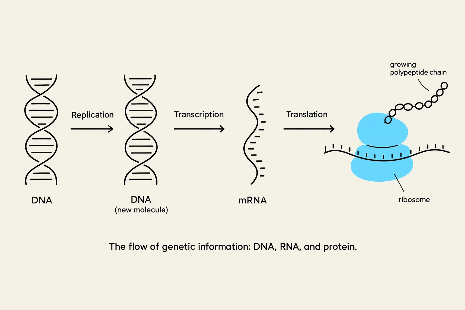
The flow of genetic information: DNA, RNA, and protein.
The "Central Dogma" describes the flow of genetic information through three distinct processes:
1. DNA Replication (DNA → DNA)
Before a cell divides, it must copy its entire genome so that daughter cells receive the necessary instructions. This process is semiconservative, meaning each new DNA molecule consists of one original "old" strand and one newly synthesized "new" strand.
The enzyme DNA polymerase is responsible for catalyzing the addition of nucleotides to a growing strand. It reads the template strand in a 3′→5′ direction and synthesizes the new strand in a 5′→3′ direction. Because DNA polymerase cannot start from scratch, it requires a primer to provide a free $3'$ end.
2. Transcription (DNA → RNA)
Transcription is the process of copying a specific segment of DNA (a gene) into an RNA molecule. Unlike replication, which copies the whole genome, transcription only copies selected regions. The enzyme RNA polymerase facilitates this, building an RNA strand from ribonucleotides.
In eukaryotic cells, the initial product is pre-mRNA, which must undergo RNA processing before it can be used for protein synthesis:
Splicing: The removal of non-coding regions called introns and the joining of coding regions called exons.
Modifications: The addition of a $5'$ cap and a poly-A tail to the ends of the molecule to ensure stability and proper use by the cell.
3. Translation (mRNA → Protein)
Translation is the final step where the information in the mRNA message is used to assemble amino acids into a polypeptide (protein). This occurs at the ribosome.
The "language" of translation is the genetic code, which uses three-base sequences called codons to specify particular amino acids.
tRNA and Anticodons: Transfer RNA (tRNA) molecules act as adapters; they carry a specific amino acid and possess an anticodon that pairs with the complementary mRNA codon.
Degeneracy: The genetic code is degenerate, meaning that multiple different codons can specify the same amino acid.
Start and Stop: The codon AUG typically signals the start of translation (specifying methionine), while specific "stop" codons signal the end of the polypeptide chain.
Laboratory Applications: PCR and Electrophoresis
Polymerase Chain Reaction (PCR)
PCR is a technique used to amplify (make many copies of) a specific DNA target in a test tube. It involves three repeating temperature-dependent steps:
Denaturation (≈95∘C): Heat separates the double-stranded DNA into single strands.
Annealing (50–75∘C): Temperature is lowered to allow primers to bind to their complementary sequences.
Extension (≈72∘C): DNA polymerase adds dNTPs to the primers to synthesize new DNA.
PCR is widely used in forensics (DNA profiling), parentage testing, and identifying pathogens.
Electrophoresis: Separating Molecules
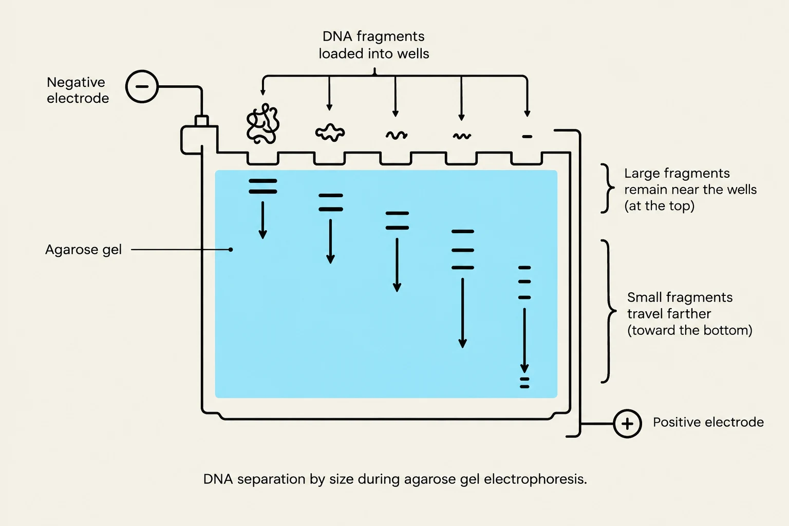
Diagram illustrating DNA separation by size using agarose gel electrophoresis, where larger fragments move slower and remain closer to the wells, while smaller fragments move faster and travel further down the gel towards the positive electrode.
Electrophoresis is used to separate DNA, RNA, or proteins based on their physical properties by moving them through a gel matrix using an electric field.
DNA/RNA Separation: Because the phosphate backbone makes nucleic acids negatively charged, they migrate toward the positive electrode. In an agarose gel, separation is primarily based on size: smaller fragments move through the pores more easily and travel farther than larger fragments.
Protein Separation: Proteins are more complex because they vary in charge and shape.
Native PAGE separates proteins based on a combination of size, shape, and charge.
SDS-PAGE uses a detergent (SDS) to unfold proteins and give them a uniform negative charge, allowing separation based almost entirely on size.
Isoelectric Focusing (IEF) separates proteins based on their isoelectric point (pI), the specific pH at which the protein has zero net charge.
Exam Tip: Comparing DNA vs. Protein Electrophoresis
Remember that while DNA gels (like agarose) almost always separate by fragment length, protein separation depends heavily on the method used (Native vs. SDS-PAGE vs. IEF).
Keep reading for free.
Create an account to finish this reading and make it part of your study routine.