An in-depth exploration of the liver's microanatomy, including its cellular composition, vascular supply, and the functional zonation of hepatic lobules. This content details hepatocytes, Kupffer cells, Ito cells, and the roles of the portal triad and hepatic acinus in liver function and metabolic gradients.
The liver is the largest internal organ in the body, performing a vast array of essential metabolic, immune, and secretory functions. In adult carnivores, it typically accounts for 3% to 4% of total body weight, whereas in large herbivores, it may represent only about 1%. Despite variations in size and lobe morphology across different species, the fundamental microanatomical architecture remains remarkably consistent among domestic animals. Understanding the liver requires an integrated view of its cellular components, its complex vascular supply, and the specialized way its tissue is organized to facilitate metabolic gradients.
Cellular Composition and the Microenvironment
The liver tissue is composed of several specialized cell types that work in concert to maintain homeostasis. The "workhorses" of the organ are the hepatocytes, which make up approximately 75% of the liver mass. These polygonal epithelial cells are arranged in cords or plates and are responsible for critical processes including detoxification, deamination (converting amino acids to urea), bile production, gluconeogenesis, and the storage of energy as glycogen and triglycerides.
Surrounding the hepatocytes and within the vascular channels are other vital cells:
Kupffer cells: These are resident, fixed macrophages located within the hepatic sinusoids. They serve an immune function by removing aged blood cells, pathogens, and certain toxins through phagocytosis.
Ito cells (Hepatic Stellate Cells): Located within the perisinusoidal space (space of Disse), these cells are characterized by large, round lipid vacuoles used for Vitamin A metabolism. They also play a crucial role in regulating sinusoidal blood flow.
Pit cells: These are resident granular leukocytes (Natural Killer cells) found within the sinusoids.
Oval cells: These serve as pluripotent stem cells and are typically found within the biliary tree.
Clinical Correlation: Hepatic Fibrosis
During hepatic injury, Ito/stellate cells undergo a dramatic transformation. They lose their Vitamin A content and acquire a myofibroblast-like phenotype, leading to the excessive production of collagen and other extracellular matrix components. This process results in hepatic fibrosis.
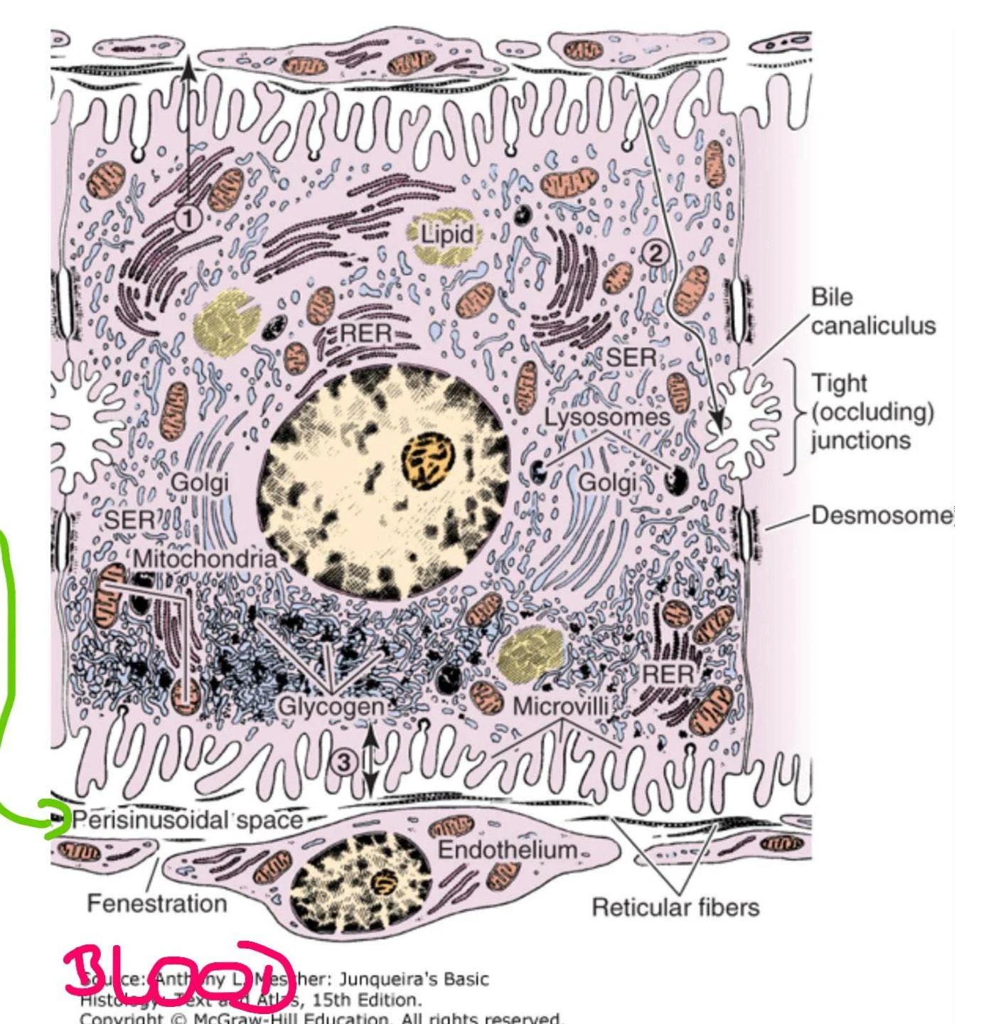
The perisinusoidal space (space of Disse) is a critical microanatomical feature located between the hepatocytes and the endothelial cells of the sinusoids. It serves as the primary site of exchange between the blood in the sinusoids and the hepatocytes.
Diagram illustrating the structure of a liver cell, showing organelles such as mitochondria, Golgi apparatus, endoplasmic reticulum (RER and SER), lysosomes, and glycogen granules. It also depicts microvilli, tight junctions, desmosomes, bile canaliculi, fenestrations in the endothelium, and reticular fibers.
Hepatic Circulation and the Portal Triad
The liver possesses a unique dual blood supply that ensures it receives both nutrient-rich blood from the digestive tract and oxygenated blood from the systemic circulation.
Portal Vein: Drains blood from the digestive tract, pancreas, and spleen, providing approximately 70% to 80% of the total afferent hepatic blood flow.
Hepatic Artery: Provides the remaining 20% to 30% of the blood supply, which is highly oxygenated.
Blood flows from the portal areas through the hepatic sinusoids—specialized, fenestrated capillaries—and eventually drains into the central vein. From the central vein, the blood exits via the hepatic veins to enter the caudal vena cava.
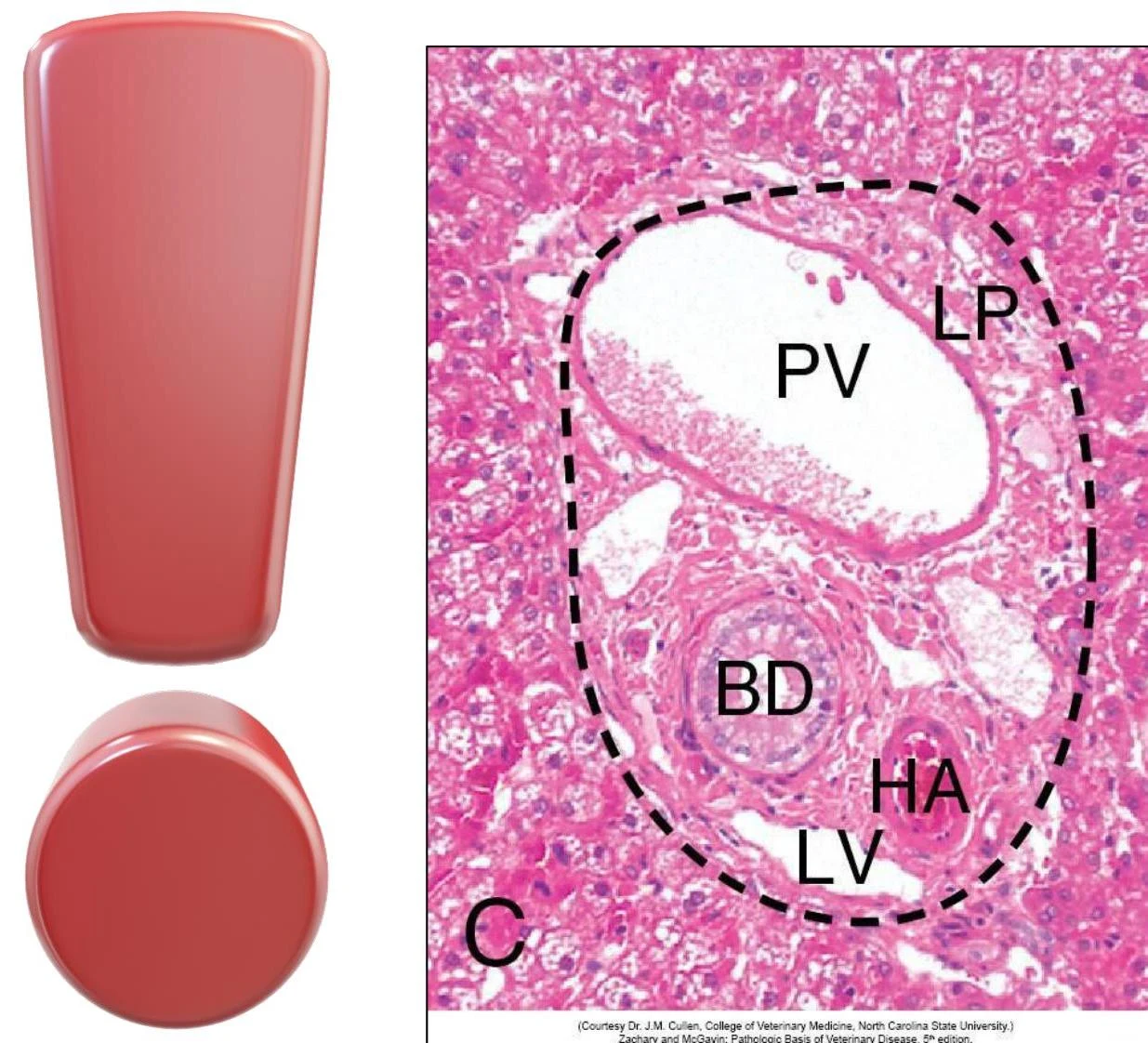
The structural units of the liver are organized around the portal triad, a cluster of vessels found at the periphery of the lobule. Each triad contains:
A branch of the hepatic portal vein.
A branch of the hepatic artery.
A branch of the bile duct.
These structures are often surrounded by a collagenous extracellular matrix that forms a distinct border with the hepatocytes, known as the limiting plate.
This is a micrograph of a portal triad in the liver, showing the portal vein (PV), hepatic artery (HA), and bile duct (BD) surrounded by liver parenchyma (LP) and lobular vein (LV).
The Biliary Tree and Bile Secretion
While blood flows toward the central vein, bile flows in the opposite direction. Hepatocytes actively secrete bile acids into tiny potential spaces between adjacent cells called bile canaliculi. Bile acids act as detergents to assist in the intestinal absorption of fats and fat-soluble vitamins.
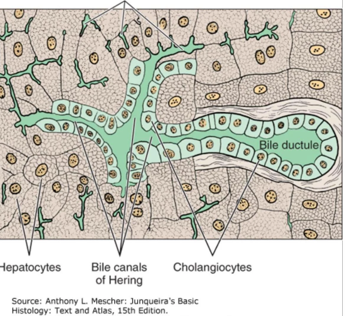
The bile canaliculi lead into larger canals and ductules lined by specialized epithelial cells called cholangiocytes. These bile ductules are located within the portal triads, forming part of the intrahepatic biliary tree.
Diagram illustrating hepatocytes, bile canals of Hering, cholangiocytes, and a bile ductule within the liver.
Functional Organization: Lobules and Zonation
To understand how the liver functions, we must distinguish between different anatomical models of organization: the classic hepatic lobule, the portal lobule, and the hepatic acinus.
Classic Hepatic Lobule: A hexagonal structure centered on the central vein. It is designed to illustrate blood flow from the portal triads at the periphery toward the center.
Portal Lobule: A triangular area centered on a portal triad, designed to illustrate the drainage of bile from hepatocytes to the bile ducts.
Hepatic Acinus: A diamond-shaped functional unit that focuses on the metabolic gradients and oxygen supply to the hepatocytes.
Hepatic Zonation
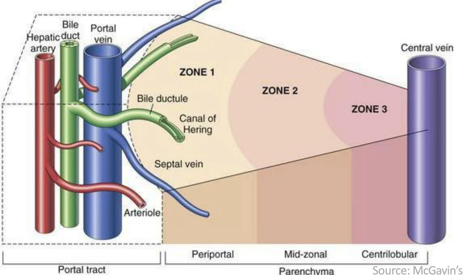
The hepatic acinus is divided into three distinct metabolic zones based on their proximity to the blood supply from the portal triad:
Zone 1 (Periportal Area): These hepatocytes are closest to the portal triad and receive the most oxygenated and nutrient-rich blood. Because they are the first to encounter incoming substances, they are more susceptible to direct-acting toxins (e.g., white phosphorus).
Zone 2 (Midzonal Area): An intermediate zone between the periportal and centrilobular areas.
Zone 3 (Centrilobular Area): These cells are located closest to the central vein. They receive blood that has already been partially depleted of oxygen and nutrients, making them highly susceptible to hypoxia. Furthermore, Zone 3 contains high levels of enzymes responsible for xenobiotic biotransformation; consequently, toxins that require metabolic activation to become harmful (such as acetaminophen) typically cause injury in this zone.
Diagram illustrating the three zones of the hepatic lobule, showing the portal tract with hepatic artery, bile duct, and portal vein, and the central vein.
Exam Tip: Patterns of Injury
If a toxin is directly injurious to cells, look for periportal (Zone 1) damage. If a toxin requires bioactivation (metabolism) to become toxic, look for centrilobular (Zone 3) damage.
In summary, the liver's microanatomy is a highly organized system where blood and bile flow in opposing directions through a specialized cellular landscape, ensuring efficient metabolism and defense.
Keep reading for free.
Create an account to finish this reading and make it part of your study routine.