Explores the roles of Major Histocompatibility Complex (MHC) molecules and T-Cell Receptors (TCRs) in distinguishing self from non-self and orchestrating immune responses through antigen presentation.
The immune system's ability to distinguish "self" from "non-self" is not a matter of simple recognition, but a complex biochemical dialogue. At the heart of this dialogue are two critical components: the Major Histocompatibility Complex (MHC) and the T-Cell Receptor (TCR). While antibodies can recognize free-floating antigens in the blood, T-cells require a more formal introduction. They cannot "see" an antigen unless it is processed and "presented" on the surface of another cell via MHC molecules. This article explores how these molecules work together to orchestrate both humoral and cell-mediated immunity.
The Major Histocompatibility Complex (MHC)
The Major Histocompatibility Complex (MHC) is a set of genes that encode for proteins located on the surface of antigen-presenting cells. These proteins act as "display cases" for small fragments of proteins (peptides) derived from pathogens. In humans, these MHC molecules are specifically referred to as Human Leukocyte Antigens (HLA).
The diversity of the MHC is one of the most striking features of the human genome. MHC genes are highly polymorphic, meaning there are many different versions (alleles) of these genes within the population. These alleles are inherited in sets called haplotypes—one set from each parent. Because these genes are co-dominantly expressed, the products of both the maternal and paternal sets are displayed on the cell surface simultaneously.
Exam Tip: If a student is asked why different individuals respond differently to the same pathogen, the answer often lies in their unique MHC haplotypes. A "hole in the repertoire" occurs if an individual's specific MHC molecules are unable to bind and present a particular antigen, leaving them vulnerable to that specific threat.
This diagram illustrates co-dominant expression of MHC haplotypes. A child inherits one MHC haplotype from each parent, and both maternal and paternal MHC molecules are expressed on the cell surface.
MHC Class I vs. MHC Class II
Not all MHC molecules are created equal. They are divided into two primary functional classes that serve different roles in the immune response.
MHC Class I: The Internal Monitor
MHC Class I molecules are found on all nucleated cells in the body. Their primary job is to report on the internal health of the cell. They present endogenous antigens—proteins that are synthesized inside the cell. This includes normal self-proteins, but more importantly, it includes viral proteins or tumor proteins produced by an infected or cancerous cell.
When a cell presents a foreign peptide via MHC Class I, it acts as a signal to cytotoxic T cells (CD8+ T cells), essentially saying, "I am infected; please destroy me". These molecules are encoded by the A, B, and C regions of the MHC.
MHC Class II: The External Scout
In contrast, MHC Class II molecules are restricted to specialized antigen-presenting cells (APCs), such as macrophages, monocytes, and B cells. Their role is to monitor the extracellular environment by presenting exogenous antigens—pathogens that have been swallowed (phagocytosed) from outside the cell.
MHC Class II molecules are encoded by the DR, DP, and DQ regions. When they present an antigen, they activate helper T cells (CD4+ T cells), which then coordinate the broader immune response, including helping B cells produce antibodies.
Feature
MHC Class I
MHC Class II
Cellular Distribution
All nucleated cells
Professional Antigen-Presenting Cells (APCs)
Antigen Type
Endogenous (Internal)
Exogenous (External/Phagocytosed)
Target T-Cell
Cytotoxic T cells (CD8+)
Helper T cells (CD4+)
Genetic Regions
A, B, C
DR, DP, DQ
T-Cell Receptors (TCRs) and Signaling
If the MHC is the "display case," the T-Cell Receptor (TCR) is the "eye" of the T-cell. T-cells use TCRs to recognize the specific peptide-MHC complex.
Structure and Diversity
TCRs are composed of heterodimers—two different protein chains linked together by a single disulfide bond. There are two main types of TCR heterodimers:
αβ (alpha/beta) receptors: The most common type in humans.
γδ (gamma/delta) receptors: A less common type.
The ability of T-cells to recognize an almost infinite variety of pathogens is due to V-J diversity. During T-cell development, different segments of the gene (Variable and Joining segments) are rearranged to create unique receptors. For example, the αβ receptor can generate approximately 2,500 combinations through this process.
Note the Difference: Unlike B-cell receptors (immunoglobulins), TCRs do not undergo somatic mutations to increase diversity, and they do not rearrange their constant region genes. Furthermore, TCRs are found only on the cell surface.
The Role of CD3
A TCR cannot communicate with the rest of the cell on its own. It is associated with a complex of proteins called CD3 (including γ, δ, and ϵ subunits) and the ζ (zeta) chain. When the TCR binds to an antigen-MHC complex, the CD3 complex acts as the signaling machinery, transmitting the message into the cell's interior to trigger an immune response. If CD3 expression is low, the immune response will be impaired because the cell cannot "hear" the signal that an antigen has been found.
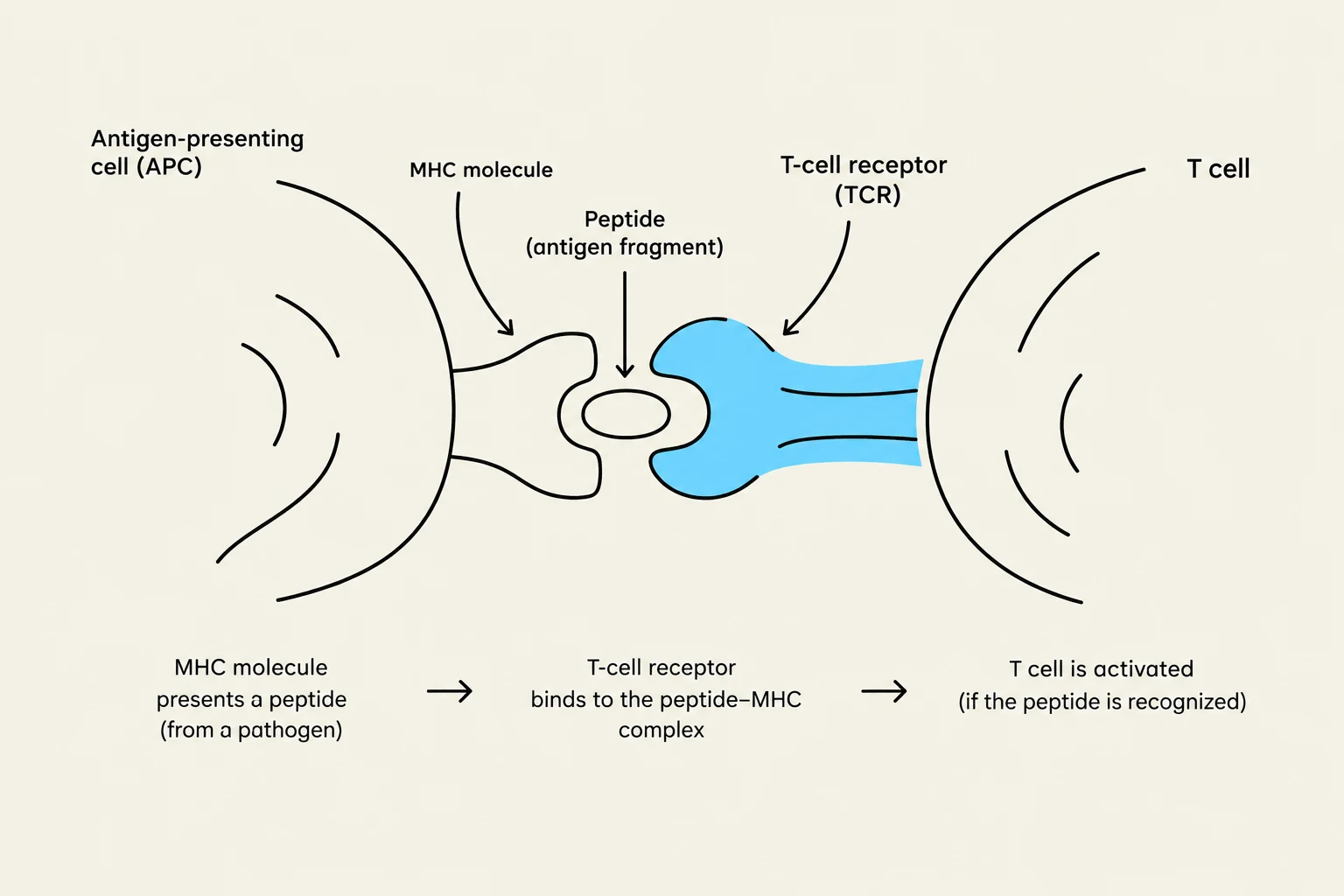
This diagram illustrates the interaction between an antigen-presenting cell (APC) and a T cell. The APC presents a peptide fragment from a pathogen via an MHC molecule, which is then recognized by the T-cell receptor (TCR) on the T cell, leading to T cell activation.
The Process of Antigen Presentation
The journey from a pathogen to an activated T-cell involves several highly regulated steps:
Internalization: An APC (like a macrophage) encounters an exogenous antigen and phagocytoses it.
Processing: The antigen is broken down biochemically into smaller peptide fragments within endosomes or lysosomes.
MHC Loading:
Class I pathway: Peptides from the endoplasmic reticulum (ER) are brought in via transporters (TAP-1 and TAP-2) to meet MHC Class I molecules.
Class II pathway: MHC Class II molecules are synthesized in the ER with an invariant chain (Ii) attached to prevent them from binding the wrong peptides. They travel to the endosomal compartment, where the invariant chain is removed, allowing the exogenous peptide to bind.
Presentation: The peptide-MHC complex is transported to the cell surface for T-cell inspection.
The efficiency of this process can be modulated. For instance, when activated T-cells release Interferon-γ, it can cause an increase in the expression of MHC Class II molecules on monocytes and macrophages, enhancing the immune response. However, excessive expression of these molecules is sometimes linked to autoimmune diseases.
Keep reading for free.
Create an account to finish this reading and make it part of your study routine.